Ko143 BCRP inhibitor Probes ABCG2 Function at the Blood-Brain Barrier in Alzheimer Models
Copy code to add this image to your page
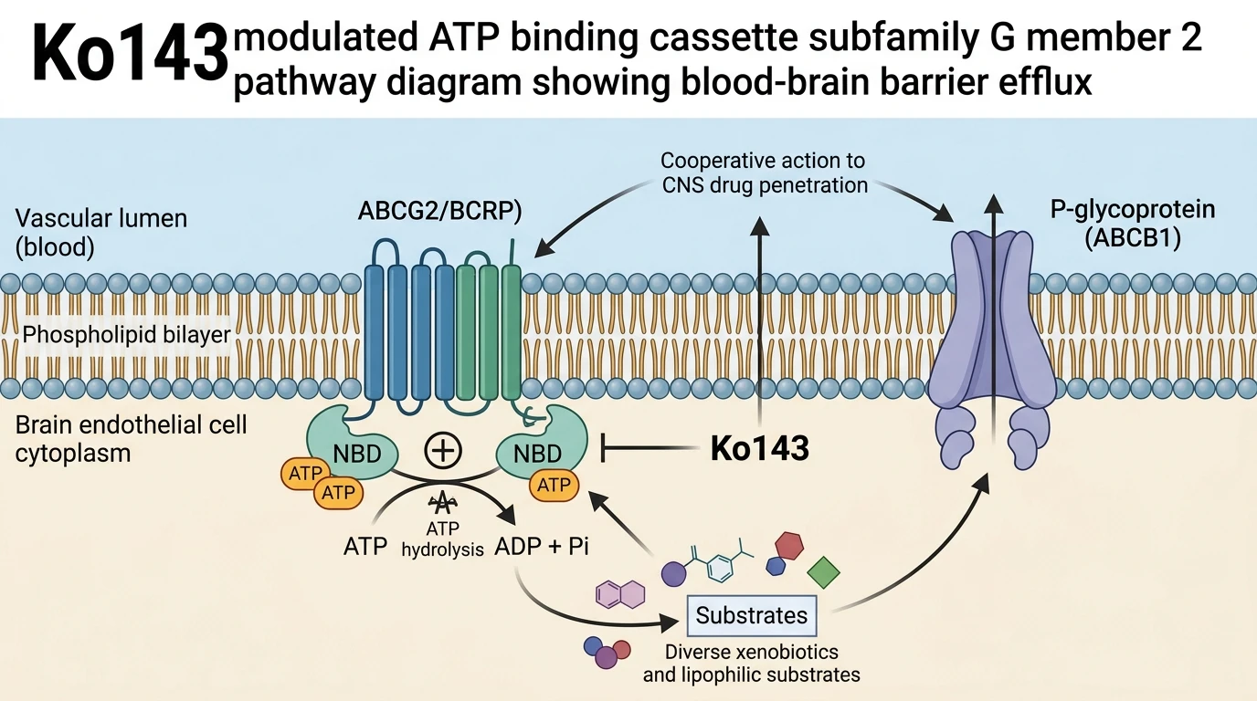
ATP-binding cassette subfamily G member 2 (ABCG2), historically recognized as breast cancer resistance protein (BCRP), functions as a primary ATP-dependent efflux pump located at the human and rodent blood-brain barrier. Localized predominantly to the luminal plasma membrane of brain capillary endothelial cells, ABCG2 utilizes energy derived from ATP hydrolysis to export structurally diverse lipophilic cations, organic anions, and xenobiotics back into the circulating blood stream. Operating in tandem with P-glycoprotein (ABCB1), ABCG2 establishes a rigorous biochemical gatekeeper system. This coordinated efflux apparatus significantly restricts the central nervous system entry of numerous small-molecule drugs, including targeted tyrosine kinase inhibitors and direct-acting neurotherapeutics.
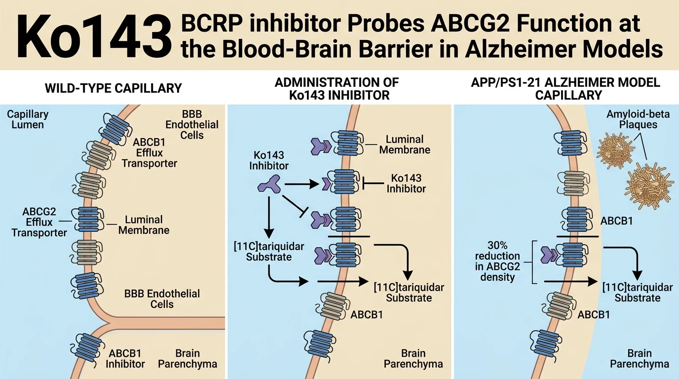
Pathological states such as Alzheimer's disease induce progressive structural and biochemical alterations across the cerebral microvasculature. In the APP/PS1-21 transgenic mouse model, which expresses human amyloid precursor protein and presenilin 1 mutations, extensive amyloid-beta deposition correlates with neurovascular unit disruption. Quantitative immunochemistry and targeted proteomic analyses demonstrate a statistically significant 29% to 37% reduction in cerebral ABCG2 microvessel protein abundance in APP/PS1-21 mice compared with age-matched wild-type controls. This substantial decrease in transporter density prompted hypotheses that central brain accumulation of dual ABCB1/ABCG2 substrates would rise during neurodegenerative disease progression.
Evaluating small-molecule transport kinetics across the blood-brain barrier in vivo demands highly selective chemical inhibitors. The synthetic compound Ko143 functions as a potent, highly selective ABCG2 antagonist, displaying nanomolar inhibitory activity while demonstrating negligible effects on ABCB1 or multidrug resistance-associated proteins at active working concentrations. Utilizing non-invasive positron emission tomography substrate imaging studies with the dual ABCB1/ABCG2 radiotracers [11C]tariquidar and [11C]erlotinib, researchers evaluated whether down-regulated microvascular ABCG2 expression translates directly into altered central drug exposure in transgenic models.
Baseline radiotracer distribution studies revealed an unexpected outcome. Despite the clear 29% to 37% reduction in physical microvascular ABCG2 expression, brain penetration of both [11C]tariquidar and [11C]erlotinib remained identical between wild-type and APP/PS1-21 mice under uninhibited conditions. Quantitative regional volume of distribution (VT) values showed no significant divergence across cerebral cortex, hippocampus, or thalamic regions between genotypes. Basal endothelial efflux transport capacity appears fully capable of sustaining maximum drug clearance even when transporter density is reduced by one-third.
To challenge the system and assess functional reserve capacity, investigators administered partial pharmacological inhibition using the Ko143 BCRP inhibitor at a sub-maximal dose of 5 mg/kg via intraperitoneal injection. Acute dosing with Ko143 rapidly attenuated ABCG2-mediated transport functionality across the cerebrovascular endothelium. Serial PET imaging demonstrated that partial transport block yielded a prominent, immediate surge in central brain uptake of [11C]tariquidar. Crucially, the relative percentage increase in brain radiotracer accumulation was identical in wild-type mice and APP/PS1-21 Alzheimer's model animals.
These empirical observations define vital functional boundaries regarding blood-brain barrier transport physiology during neurodegeneration. A moderate decrease of approximately 30% in microvascular ABCG2 abundance does not produce functional barrier failure in vivo. Cerebral microvessels possess a broad excess catalytic capacity for substrate clearance. Consequently, moderate reductions in ABCG2 expression observed in neurodegenerative animal models do not alter central drug exposure, reflecting robust baseline reserve capacity and overlapping functional protection provided by concurrent ABCB1 efflux.
In addition, the overlapping substrate specificity between ABCB1 and ABCG2 provides a compensatory safety mechanism at the capillary interface. When ABCG2 molecules are reduced in density or partially occupied, ABCB1 actively accommodates substrate flux, limiting net parenchymal entry. Experimental co-inhibition and targeted substrate kinetics indicate that both transporters operate in a highly cooperative thermodynamic equilibrium. Consequently, therapeutic strategies aimed at delivering neuropharmaceuticals across an impaired blood-brain barrier cannot rely solely on disease-induced transporter down-regulation. Targeted inhibition using validated tools like the Ko143 BCRP inhibitor remains necessary to produce measurable alterations in brain tissue concentrations.
Functional Evaluation of Cerebrovascular Efflux Transporters in Transgenic Models
Copy code to add this image to your page
ABCG2 active efflux at the blood-brain barrier maintains high reserve capacity against localized microvascular loss. In APP/PS1-21 mice, a 29–37% reduction in cerebral ABCG2 abundance produces no shift in baseline drug distribution. Pharmacological challenge using Ko143 (5 mg/kg) increases brain uptake of dual ABCB1/ABCG2 substrates ([11C]tariquidar) symmetrically across wild-type and transgenic strains. This functional redundancy buffers central nervous system pharmacokinetics against moderate transporter down-regulation.
Microvascular ABCG2 Depletion Fails to Alter Baseline Brain Penetration
Quantitative immunochemistry confirmed a 29% to 37% reduction of ABCG2 in APP/PS1-21 microvessels. Baseline PET imaging with [11C]tariquidar and [11C]erlotinib demonstrated equivalent radiotracer distribution across both strains. Cerebral volume of distribution metrics remained unchanged. Physiological efflux capacity exceeds basal clearance requirements at the blood-brain barrier. Consequently, partial loss of transporter expression does not elevate substrate brain accumulation.
Ko143 Administration Identifies Substantial Transport Reserve Capacity
Systemic treatment with the Ko143 BCRP inhibitor at 5 mg/kg unmasked latent efflux dynamics in vivo. Acute target engagement induced a marked surge in central [11C]tariquidar accumulation. Radiotracer brain uptake values rose symmetrically in wild-type and transgenic animals. Absolute transport capacity remains functionally redundant under physiological challenge. Pharmacological intervention with the Ko143 BCRP inhibitor confirmed that remaining microvascular ABCG2 proteins maintain adequate clearance velocities.