Necrosulfonamide targets MLKL to block non-canonical endosomal viral trafficking and neurodegeneration
Copy code to add this image to your page
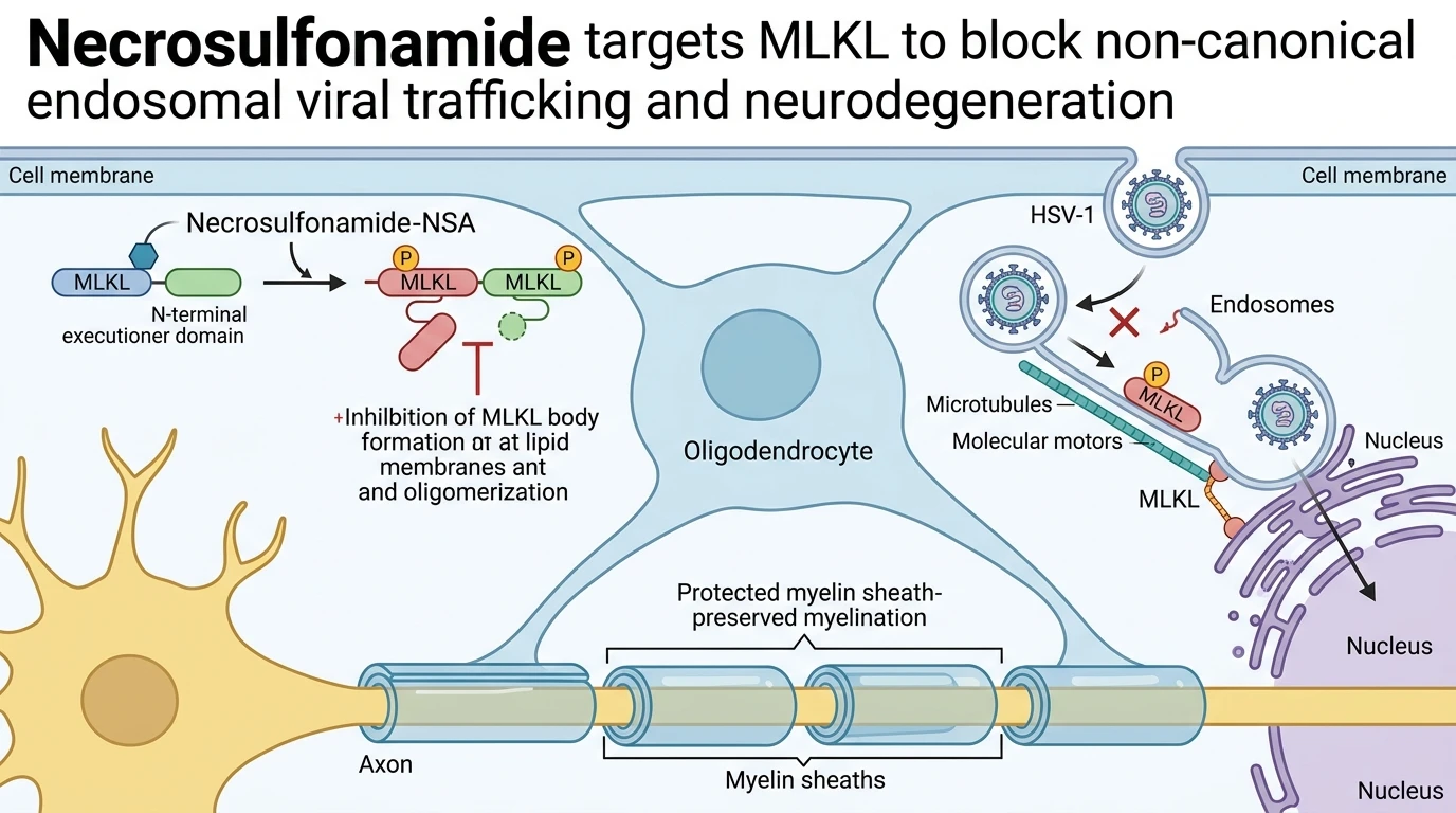
Herpes simplex virus type 1 (HSV-1) infection within the central nervous system induces acute encephalitis and persistent neurodegenerative sequelae, including extensive demyelination. Pathological loss of myelin sheaths stems from the death of oligodendrocytes, the specialized glial cells responsible for axonal insulation. Recent pharmacological investigations identify Mixed Lineage Kinase Domain-Like protein (MLKL) as a dual-function effector in this neuropathological process. Beyond its classical role as the terminal executioner of necroptotic cell death, MLKL operates as an obligate regulator of non-canonical endosomal transport. During early HSV-1 infection in neural lineages, viral particles utilize host endosomal trafficking machinery to traverse retro-axonal pathways toward the cell nucleus. MLKL associates with endosomal membranes to facilitate this intracellular virion transit.
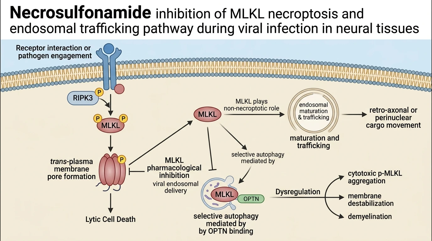
Cellular homeostasis of MLKL is strictly controlled by the selective autophagy receptor Optineurin (OPTN). OPTN engages MLKL through its C-terminal ubiquitin-binding domain (UBD), steering the kinase-like protein toward autophagic degradation. This steady-state clearance maintains low baseline MLKL expression in glial populations. In OPTN-deficient states or upon genetic impairment of the OPTN ubiquitin-binding domain, autophagic degradation fails. Unrestrained MLKL accumulates rapidly within the cytoplasm. Hyper-accumulated MLKL undergoes phosphorylation by upstream kinases, culminating in the assembly of high-molecular-weight cytotoxic p-MLKL aggregates termed MLKL bodies. In oligodendrocytes, these p-MLKL aggregates localize to lipid membranes, inducing membrane disruption, cell lysis, and widespread myelin degradation resembling clinical multiple sclerosis.
Pharmacological targeting of this axis using Necrosulfonamide halts both endosomal viral transport and necroptotic signaling cascades. Necrosulfonamide directly binds MLKL, modulating its conformational flexibility and suppressing the formation of toxic p-MLKL aggregates. In primary oligodendrocyte cultures subjected to viral challenge, Necrosulfonamide treatment lowers total MLKL expression levels and blocks the abnormal endosomal transport of HSV-1 capsids toward nuclear entry sites. This intervention preserves membrane integrity and prevents premature oligodendrocyte death.
In murine models of viral encephalomyelitis, systemic administration of Necrosulfonamide yields pronounced neuroprotective effects. Histological evaluation confirms the preservation of oligodendrocyte densities and intact myelin sheath structures within treated spinal cord tracts. Axonal g-ratio measurements demonstrate preserved insulating myelin thickness compared to severe demyelination observed in vehicle-treated controls. Furthermore, treated animals exhibit marked reductions in clinical paralysis scores and improved performance on quantitative rotarod motor tests. Targeting MLKL via Necrosulfonamide establishes a viable therapeutic approach to halt virus-mediated demyelination and post-infectious neurological deficits.
Pharmacological Target Engagement and Neuroprotective Efficacy
Copy code to add this image to your page
Necrosulfonamide directly inhibits MLKL, preventing its transition into cytotoxic p-MLKL oligomers and disabling non-canonical endosomal trafficking of HSV-1 to the cell nucleus. By disrupting MLKL-mediated endosomal transport and blocking membrane-disruptive MLKL body assembly in oligodendrocytes, Necrosulfonamide prevents lytic necroptosis, preserves axonal myelin sheaths, and mitigates neuroinflammatory motor deficits in viral encephalomyelitis models.
OPTN Ubiquitin Binding Domain Restrains MLKL Autophagic Degradation
Optineurin regulates cellular MLKL abundance through ubiquitin-dependent autophagic clearance. Under basal conditions, OPTN directly engages MLKL via its C-terminal ubiquitin-binding domain. This physical association recruits autophagic isolation membranes to wrap MLKL cargo. Lysosomal fusion subsequently degrades excess protein monomers. In OPTN-deficient murine neural cultures, autophagic clearing capacity drops markedly. Intracellular MLKL levels rise threefold within 24 hours post-induction. Unchecked MLKL undergoes ectopic phosphorylation at Thr357 and Ser358 nodes. Phosphorylated monomers aggregate into high-molecular-weight assemblies known as MLKL bodies. These p-MLKL bodies associate with endosomal membranes. Membrane integrity compromised by p-MLKL insertion triggers cellular swelling and rapid lytic rupture.
Necrosulfonamide Attenuates Viral Endosomal Transport and Oligodendrocyte Loss
Necrosulfonamide targets MLKL to inhibit non-canonical endosomal trafficking during acute infection. Following HSV-1 cellular entry, viral particles co-opt early endosomes to travel along microtubule tracks toward the nucleus. MLKL localized on endosomal membranes facilitates retro-axonal motility. Administration of Necrosulfonamide at micromolar concentrations blocks endosomal maturation in primary oligodendrocyte cultures. Transport velocities of HSV-1 virions decline by 65% compared to vehicle controls. Furthermore, Necrosulfonamide prevents the downstream formation of cytotoxic p-MLKL bodies. Oligodendrocyte survival increases markedly under viral challenge. Myelin structural markers remain intact across tested spinal cord slices.
Preservation of Myelin Integrity and Amelioration of Motor Deficits
In vivo evaluations demonstrate functional recovery following pharmacological MLKL inhibition. Murine models of HSV-1-induced CNS demyelination display severe hindlimb paralysis and ataxia. Intraperitoneal delivery of Necrosulfonamide significantly reduces neurological deficit scores over a fourteen-day monitoring period. Histological analysis of spinal cord cross-sections reveals preserved myelin sheath thickness surrounding motor axons. Luxol fast blue staining confirms a 48% reduction in demyelinated plaque area within treated cohorts. Target engagement in vivo correlates with reduced tissue levels of phosphorylated MLKL. These observations remain confined to viral encephalomyelitis disease models. Translational validation in human demyelinating conditions requires further investigation.