GSK1016790A activates TRPV4 channels to drive spatially distinct ocular lens calcium influx
Copy code to add this image to your page
The ocular lens is an avascular structure dependent on an internal microcirculatory system to deliver nutrients, maintain transparency, and clear metabolic waste. This circulation is driven by regional differences in ion transport, fluid movement, and hydrostatic pressure gradients established primarily by sodium and calcium transport mechanisms. Among the ion channel families involved in regulating lens physiology, Transient Receptor Potential (TRP) channels operate as key cellular sensors for mechanical stress, osmotic changes, and temperature variations. Specifically, Transient Receptor Potential Vanilloid 4 (TRPV4) functions as a non-selective, calcium-permeable cation channel that translates physical and chemical cues into intracellular calcium signals.
Recent pharmacological investigations using intact rat lens models have clarified the spatial functional distribution of TRPV4 across distinct lens cell populations. Utilizing the potent small-molecule TRPV4 agonist GSK1016790A alongside the selective TRPV1 agonist capsaicin, researchers evaluated real-time intracellular calcium dynamics across three primary cellular regions: central anterior epithelial cells, equatorial epithelial cells, and differentiating fiber cells.
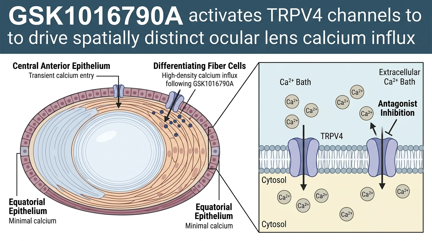
Application of GSK1016790A produced striking spatial differences in calcium mobilization. In central anterior epithelial cells, channel activation triggered a transient influx of extracellular calcium. This self-limiting response suggests tight autoregulatory mechanisms or rapid channel desensitization in the anterior epithelium. In contrast, differentiating fiber cells exhibited a significantly more robust, sustained calcium influx upon exposure to the agonist. Fiber cells form the bulk of the lens mass, and their resting intracellular calcium concentration must be rigorously controlled to prevent protein aggregation, calpain activation, and subsequent opacification. The pronounced responsiveness of differentiating fiber cells to GSK1016790A suggests that TRPV4 channels are densely expressed or highly active during fiber cell differentiation, where they likely participate in volume regulation and organelle degradation processes necessary for fiber maturation.
Interestingly, equatorial epithelial cells demonstrated minimal calcium entry following GSK1016790A administration. The equatorial region is the primary site of cell proliferation and differentiation into elongating fiber cells. The relative quiescence of TRPV4-mediated signaling in this germinative zone highlights functional compartmentalization within the lens epithelium. This non-uniform functional distribution suggests that TRPV4 activity is tuned to specific physiological tasks depending on the localized differentiation status and biomechanical demands of each cell layer.
To confirm the ionic mechanisms underlying these observed responses, experimental protocols evaluated extracellular divalent cation dependence. Omission of calcium from the bathing solution completely prevented GSK1016790A-induced calcium mobilization across all lens compartments. Re-introduction of extracellular calcium restored agonist responsiveness, confirming that the elevated intracellular signal stems from direct transmembrane calcium influx rather than internal store release from the endoplasmic or sarcoplasmic reticulum.
Pharmacological specificity was further confirmed using the selective TRPV4 antagonist HC-067047. Pre-treatment with HC-067047 completely inhibited GSK1016790A-stimulated calcium transients in both epithelial and fiber cell layers. Conversely, application of the TRPV1 agonist capsaicin failed to produce significant calcium responses under identical assay conditions. These comparative findings verify that the agonist-induced responses are specifically mediated by TRPV4 activation rather than off-target effects or non-specific TRP channel stimulation.
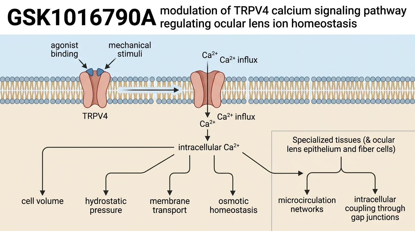
The spatial variations in TRPV4-mediated calcium entry bear directly on the mechanics of lens hydrostatic pressure and microcirculation. Current physiological models propose that surface epithelial cells generate electromotive forces that drive sodium and fluid into the lens core via extracellular spaces, while deep fiber cells clear fluid through intracellular gap junctions toward the equator and anterior surface. TRPV4 acts as an osmotic and mechanical feedback element within this loop. By modulating local calcium levels, GSK1016790A engagement influences downstream calcium-dependent effectors, including Na+/K+-ATPase activity and gap junctional conductance. Thus, TRPV4 channel activation provides a mechanism for tuning internal fluid pressure and ion homeostasis across the distinct anatomical zones of the ocular lens.
Regional Calcium Dynamics and TRPV4 Channel Modulation
Copy code to add this image to your page
GSK1016790A acts as a potent and selective agonist of TRPV4 cation channels. Upon channel binding, it induces opening of the pore, facilitating rapid influx of extracellular calcium ions down their electrochemical gradient. In intact ocular lenses, this response shows regional variation, triggering prominent calcium uptake in differentiating fiber cells and moderate transient entry in central anterior epithelial cells. This localized calcium mobilization is contingent on extracellular calcium presence and is selectively blocked by the TRPV4 antagonist HC-067047, thereby modulating lens ion transport, hydrostatic pressure, and hydration status.
Differential Calcium Influx Across Distinct Lens Zones
Central anterior epithelial cells exhibited transient calcium entry. Perfusion with GSK1016790A rapidly elevated intracellular calcium concentrations in differentiating fiber cells, whereas equatorial epithelial cells demonstrated negligible ion influx under identical experimental conditions. This marked spatial distinction points to differential channel expression or regional subunit regulation. In fiber cells, calcium accumulation reached substantial peak magnitudes within three minutes of drug application. The cellular response required external calcium. Removal of extracellular divalent cations completely abolished GSK1016790A-mediated signaling cascades across all surveyed tissue layers.
Target Specificity and Pharmacological Blockade via HC-067047
Target engagement was verified using selective pharmacological antagonists. Preincubation with the specific TRPV4 antagonist HC-067047 effectively blocked GSK1016790A-evoked calcium transients in both epithelial and fiber cell compartments. Capsaicin failed to elicit comparable responses. This contrast confirms that TRPV1 contributes minimally to the observed acute calcium flux in this rodent lens system. Ion channel inhibition remained fully reversible upon wash-out. These observations substantiate a functional role for TRPV4 in coordinating localized calcium entry required for lens microcirculation.