Napabucasin (BBI608) Inhibits STAT3 Phosphorylation and Translation Initiation in Osteosarcoma
Copy code to add this image to your page
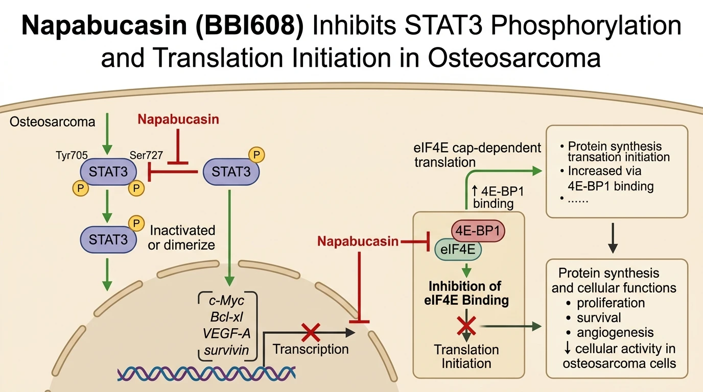
STAT3 functions as a critical oncogenic node in human osteosarcoma, orchestrating transcriptional programs that sustain cellular proliferation, survival, angiogenesis, and metastatic spread. Targeting this pathway, Napabucasin (BBI608) exhibits marked pharmacological activity against osteosarcoma cells, as demonstrated in preclinical evaluations across human osteosarcoma cell lines 143B, MG63, U2OS, and KHOS. In vitro treatment with Napabucasin suppresses cell viability in a concentration-dependent manner, concurrently reducing long-term clonogenic survival. This loss of reproductive integrity correlates with the activation of intrinsic apoptotic cascades, evidenced by the cleavage and active maturation of pro-caspase-3 into its functional heterotetramer. Substrate cleavage of poly(ADP-ribose) polymerase (PARP)—a primary substrate of active caspase-3—further confirms the execution of programmed cell death following compound exposure under tested assay conditions.
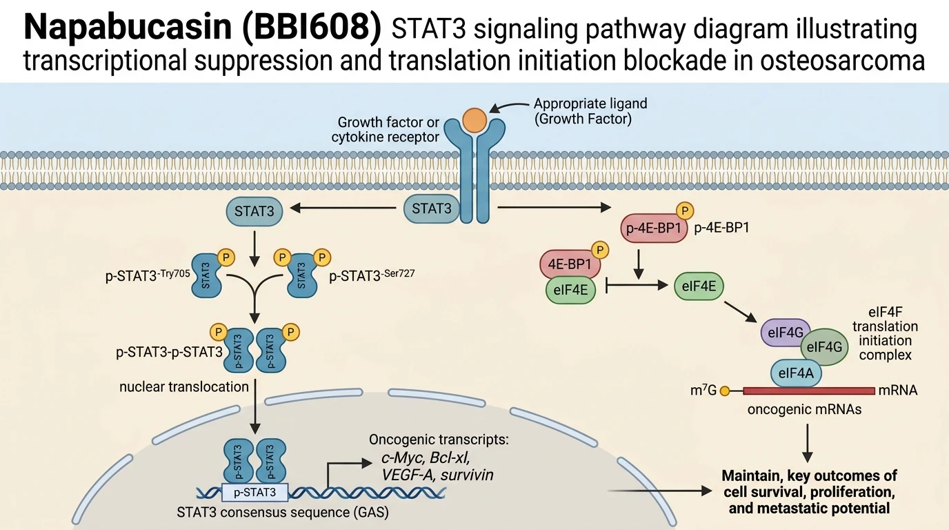
Mechanistically, STAT3 transcriptional potency depends on specific post-translational modifications. Phosphorylation at Tyr705 promotes STAT3 SH2-domain-mediated dimerization, nuclear entry, and promoter binding. Phosphorylation at Ser727 within the transactivation domain provides additional transactivation activity necessary for maximal target gene expression. Western blot and biochemical analyses reveal that Napabucasin attenuates total STAT3 protein expression while simultaneously blocking phosphorylation at both Tyr705 and Ser727 residues. Consequent to this suppression, STAT3 nuclear translocation and DNA-binding activity decline. Downstream target genes show corresponding reductions in protein abundance: the master cell cycle regulator c-Myc, the anti-apoptotic regulator Bcl-xl, the pro-angiogenic mediator VEGF-A, and the inhibitor of apoptosis survivin all exhibit marked downregulation following treatment in osteosarcoma cell models.
In addition to transcriptional blockade, Napabucasin disrupts translation initiation—a crucial bottleneck in oncogenic protein synthesis. Eukaryotic translation initiation factor 4E (eIF4E) serves as the rate-limiting cap-binding component of the eIF4F complex. Under homeostatic signaling, phosphorylation of 4E-binding protein 1 (4E-BP1) frees eIF4E to recruit eIF4G and assemble active initiation complexes. Napabucasin treatment alters this balance by increasing the binding affinity and physical association between 4E-BP1 and eIF4E, while modulating eIF4E levels. Enhanced retention of eIF4E in inactive 4E-BP1 complexes blocks assembly of the eIF4F translation machinery. Consequently, cap-dependent translation of labile, highly structured oncogenic mRNAs—including those encoding survival factors and cell cycle drivers—is selectively suppressed at the initiation step.
The in vivo efficacy of Napabucasin has been evaluated in an orthotopic tibial mouse model of osteosarcoma, which recapitulates the primary bone microenvironment and spontaneous lung metastasis observed clinically. Intratibial inoculation of 143B cells in immunocompromised mice yields rapid primary tumor expansion accompanied by aggressive cortical bone destruction and pulmonary metastasis. Administration of Napabucasin in this model led to a significant reduction in primary tibial tumor volume relative to vehicle-treated controls. Radiographic and micro-computed tomography (micro-CT) imaging demonstrated preservation of trabecular and cortical bone architecture, showing that compound administration prevents tumor-induced bone osteolysis. Furthermore, histopathological examination of lung tissue revealed a lower number of metastatic pulmonary foci.
To confirm target engagement in tissue specimens, immunohistochemical profiling of primary xenograft sections demonstrated reduced phospho-STAT3 levels alongside diminished staining for proliferation markers. The suppression of localized osteolysis suggests that STAT3 inhibition interrupts the feed-forward communication loop between osteosarcoma cells and osteoclasts within the endosteal niche. While these preclinical findings demonstrate strong activity, clinical utility requires further validation in human trial cohorts to establish therapeutic windows and identify predictive biomarkers of response.
Therapeutic Evaluation and Pharmacological Characterization
Copy code to add this image to your page
Napabucasin inhibits STAT3 transcription factor signaling by suppressing STAT3 expression and phosphorylation at Tyr705 and Ser727. This blockade downregulates transcriptional targets including c-Myc, Bcl-xl, VEGF-A, and survivin, inducing apoptosis via caspase-3 activation and PARP cleavage. Additionally, Napabucasin targets protein translation at the initiation step by modulating eIF4E and increasing 4E-BP1 association with eIF4E. In vivo, this dual activity attenuates primary osteosarcoma growth, limits lung metastases, and protects against tumor-induced bone osteolysis.
Suppression of STAT3 Tyr705 and Ser727 Phosphorylation
STAT3 activation in osteosarcoma relies on dual-site phosphorylation. Napabucasin disrupts this regulatory axis. Treatment of 143B and MG63 osteosarcoma cell lines with Napabucasin at micromolar concentrations resulted in target engagement within six hours. Western blot assays confirmed complete loss of Tyr705 phosphorylation; Ser727 phosphorylation decreased in parallel. Direct suppression of STAT3 protein levels was also detected. Consequently, transcript levels of c-Myc, Bcl-xl, VEGF-A, and survivin decreased substantially. Cellular viability dropped sharply following target downregulation. Flow cytometry confirmed a sharp increase in annexin V-positive apoptotic populations. Proteolytic cleavage of caspase-3 and PARP matched the kinetics of STAT3 suppression. These data demonstrate that target engagement directly triggers apoptotic signaling in vitro.
Blockade of eIF4E Cap Dependent Translation Initiation Complex
Translation initiation regulates short-lived oncogenic proteins in malignant bone tumors. Napabucasin targets this rate-limiting translational checkpoint. Immunoprecipitation assays revealed increased physical association between 4E-BP1 and eIF4E following exposure. This association isolated eIF4E from eIF4G binding. Assembly of the functional eIF4F complex was prevented. Cap-dependent translation rates declined rapidly as a consequence. Interestingly, total eIF4E levels were moderately altered, whereas 4E-BP1 hypophosphorylation stabilized the inhibitory complex. This translational arrest complements direct STAT3 transcriptional inhibition. Protein levels of short-half-life survival factors fell faster than predicted by mRNA decay rates alone. This dual mechanism prevents adaptive resistance arising from single-node signaling blockade in these cellular contexts.
Reduction of Tibial Tumor Burden and Lung Metastasis
Orthotopic tibial xenograft models provide a rigorous system for evaluating local and systemic efficacy. Mice bearing 143B cell xenografts received daily oral administration of Napabucasin. Primary tumor growth in the tibia slowed dramatically compared to control cohorts. Micro-CT imaging revealed marked preservation of cortical bone integrity. Osteoclast-mediated bone osteolysis was attenuated significantly. Systemic disease progression was similarly altered; lung tissue analysis demonstrated reduced metastatic lesion counts. Histological staining confirmed lower mitotic indices and reduced Ki-67 positivity in treated tissues. No signs of severe systemic toxicity or body weight loss were observed during treatment. These findings highlight the potential of targeting STAT3 and translation initiation in osteosarcoma models.