MSA-2 MSA2 STING Agonism Reprograms Macrophages to Overcome Osimertinib Resistance in EGFR-Mutant Lung Cancer
Copy code to add this image to your page
Osimertinib, a third-generation irreversible epidermal growth factor receptor (EGFR) tyrosine kinase inhibitor, demonstrates substantial clinical activity in non-small cell lung cancer harboring activating EGFR mutations (L858R and exon 19 deletions) as well as the T790M resistance mutation. Despite initial high objective response rates, acquired resistance remains a persistent clinical obstacle. Mechanistic analyses reveal that osimertinib-elicited tumor regression relies critically on intact host immunity, specifically requiring functional CD8+ T cell effector responses. Over time, sustained EGFR inhibition triggers compensatory alterations within the tumor microenvironment. Advanced lesions establish an immunosuppressive niche enriched with M2-like tumor-associated macrophages (TAMs), which directly suppress T cell recruitment, proliferation, and cytolytic output.
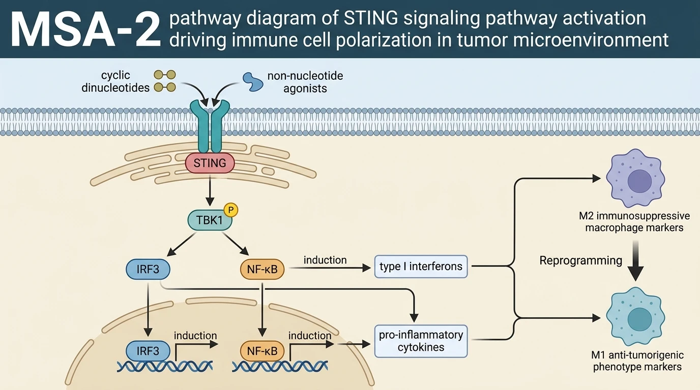
To circumvent macrophage-mediated immunosuppression, targeting innate immune sensing pathways offers a novel therapeutic paradigm. The Stimulator of Interferon Genes (STING) pathway serves as a central signaling hub in cytosolic DNA sensing. Binding of endogenous cyclic dinucleotides or synthetic small-molecule agonists to STING triggers conformational changes, leading to the recruitment and activation of TANK-binding kinase 1 (TBK1). Activated TBK1 phosphorylates the transcription factor interferon regulatory factor 3 (IRF3), promoting its nuclear translocation and driving the transcription of type I interferons (IFN-α/β) along with secondary pro-inflammatory cytokines.
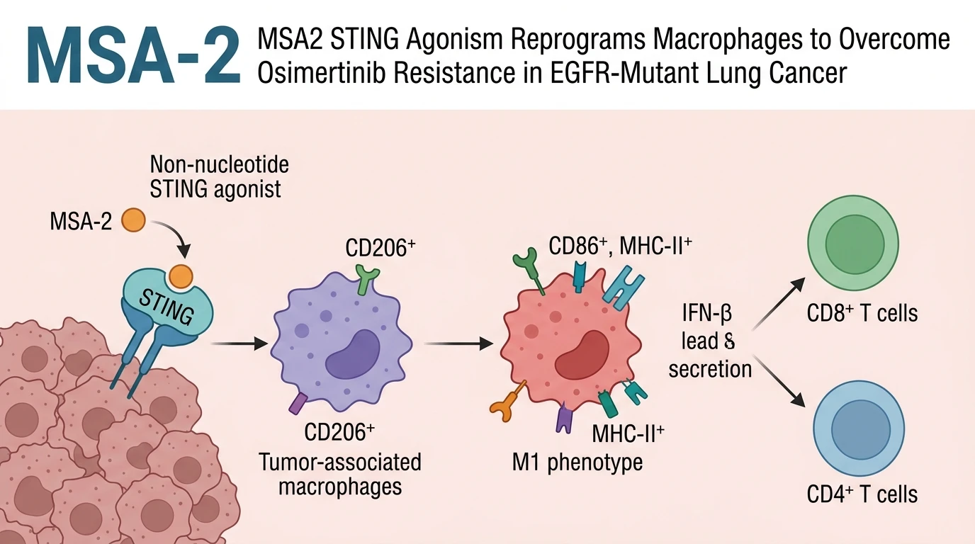
The non-nucleotide small-molecule agonist MSA-2 (also designated in preclinical literature as MSA2 small-molecule STING agonist) exhibits orally bioavailable properties and potent binding affinity for human and murine STING homodimers. Unlike canonical charged cyclic dinucleotides that suffer from rapid systemic clearance and restricted intracellular entry, MSA-2 enters target cells efficiently through passive diffusion. Within the intracellular compartment, MSA-2 stabilizes STING into its active closed dimer conformation, driving robust downstream signaling cascades.
In preclinical murine models of osimertinib-resistant EGFR-mutant lung cancer, administrative monotherapy using targeted STING agonists alters the cellular composition of the microenvironment. Specifically, treatment with MSA-2 triggers a phenotypic switch in TAMs, shifting them from a pro-tumorigenic, CD206-high M2 state to an anti-tumorigenic, CD86-high and MHC-II-high M1 state. M1-polarized macrophages release type I interferons and CXCL9/CXCL10 chemokines, which recruit CD8+ cytotoxic lymphocytes and CD4+ helper T cells into previously immunologically cold tumors.
Combining osimertinib with MSA-2 restores immune-mediated tumor clearance. In syngeneic and patient-derived xenograft models, dual inhibition reinstates intratumoral T cell proliferation, interferon-gamma expression, and granzyme B secretion. This multi-nodal mechanism addresses both cell-intrinsic oncogenic driver activity and cell-extrinsic immune escape, establishing a firm empirical rationale for combination strategies in drug-resistant EGFR-mutant non-small cell lung carcinoma.
Pharmacological Reprogramming of Macrophages and Immune Restoration
Copy code to add this image to your page
MSA-2 functions as a non-nucleotide STING agonist that targets immunosuppressive tumor microenvironments. By inducing STING activation, it drives M2-to-M1 macrophage polarization, elevating pro-inflammatory cytokines while repressing pro-tumorigenic factors. In EGFR-mutant lung carcinoma models resistant to osimertinib, this phenotypic shift reverses T cell exclusion, restoring CD8+ and CD4+ T cell infiltration, effector function, and long-term tumor control.
STING Agonism Reverses Immunosuppressive M2 Macrophage Polarization
Osimertinib resistance associates strongly with intratumoral accumulation of M2-like macrophages. These suppressive myeloid cells impede cytotoxic T cell infiltration in advanced lesions. Under tested in vitro assay conditions, administration of MSA-2 forced a rapid down-regulation of scavenger receptor CD206. Concurrently, expression of activation markers CD86 and MHC-II increased significantly. This structural shift confirms direct conversion to the M1 phenotype. Pharmacological targeting of STING neutralizing local immunosuppression within the tumor microenvironment.
Combination Therapy Restores Intratumoral Cytotoxic T Cell Recruitment
Immune cell suppression limits the durability of targeted kinase inhibitors in resistant models. Within this murine xenograft model, osimertinib alone failed to sustain CD8+ T cell activity. Adding the agonist MSA2 restored CXCL9 and CXCL10 chemokine expression. Effector lymphocytes infiltrated deep into the core of treatment-resistant tumors. Tumor cell density dropped rapidly within ten days. Flow cytometry verified elevated production of interferon-gamma and granzyme B. These observations confirm that myeloid polarization is required to unblock lymphocyte cytotoxicity in refractory tumors.