JTE 013 S1P Receptor antagonist suppresses RhoA/ROCK1/Drp1 axis in renal endothelial cells
Copy code to add this image to your page
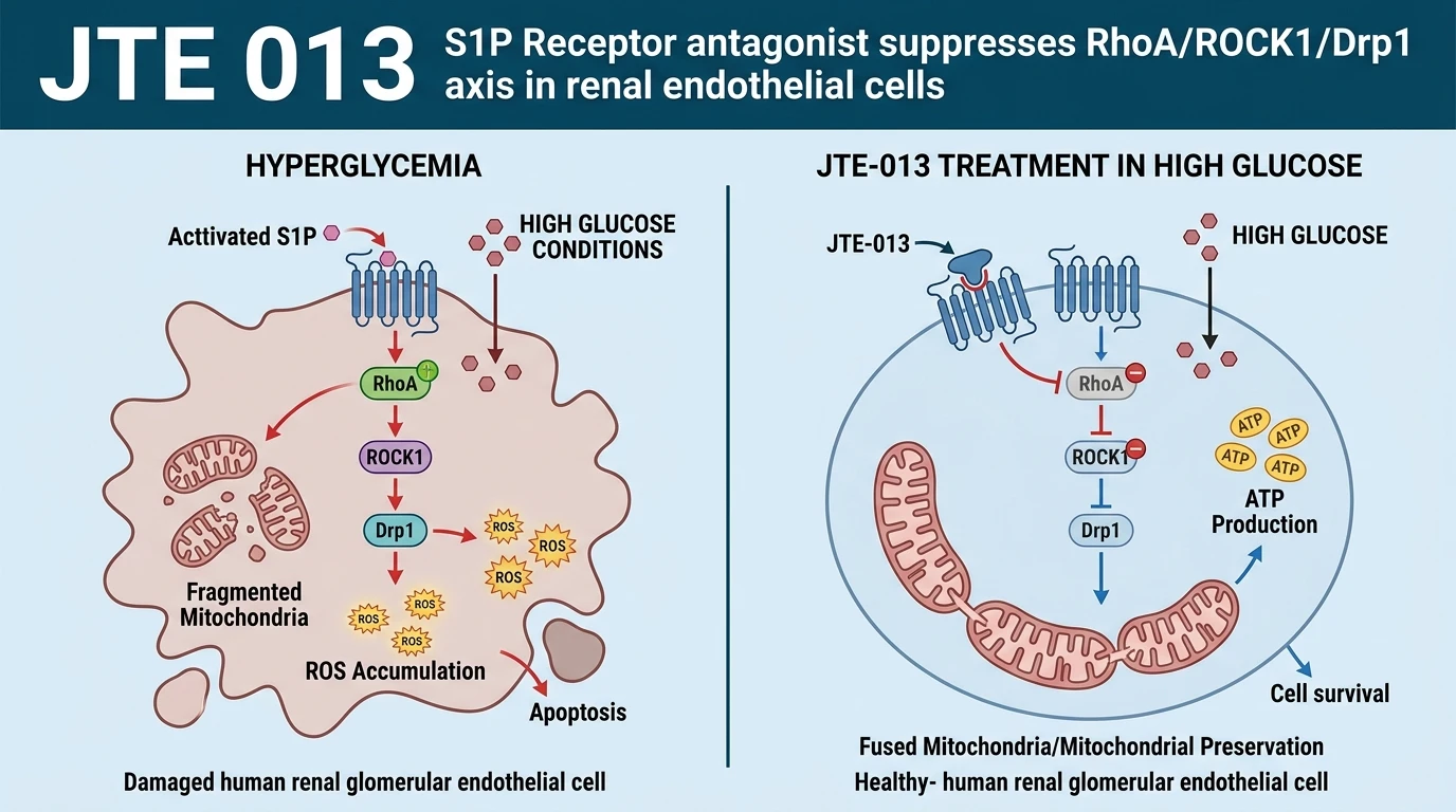
Hyperglycemic microenvironments in diabetic nephropathy induce severe metabolic dysfunction in human renal glomerular endothelial cells (HRGECs). High glucose (HG) exposure initiates intracellular biochemical perturbations that culminate in microvascular breakdown. A major driver of this pathology is aberrant mitochondrial dynamics. Healthy HRGECs rely on a balanced cycle of mitochondrial fusion and fission to maintain bioenergetic homeostasis and low basal reactive oxygen species (ROS) output. Under HG conditions, this physiological balance shifts dramatically toward excessive mitochondrial fragmentation. Fission events dominate over fusion. This structural disruption causes rapid dissipation of mitochondrial membrane potential, loss of ATP synthesis efficiency, and intracellular calcium (Ca2+) overload. Consequently, impaired endothelial cells undergo accelerated apoptosis and lose their microvascular barrier integrity and migration capacity.
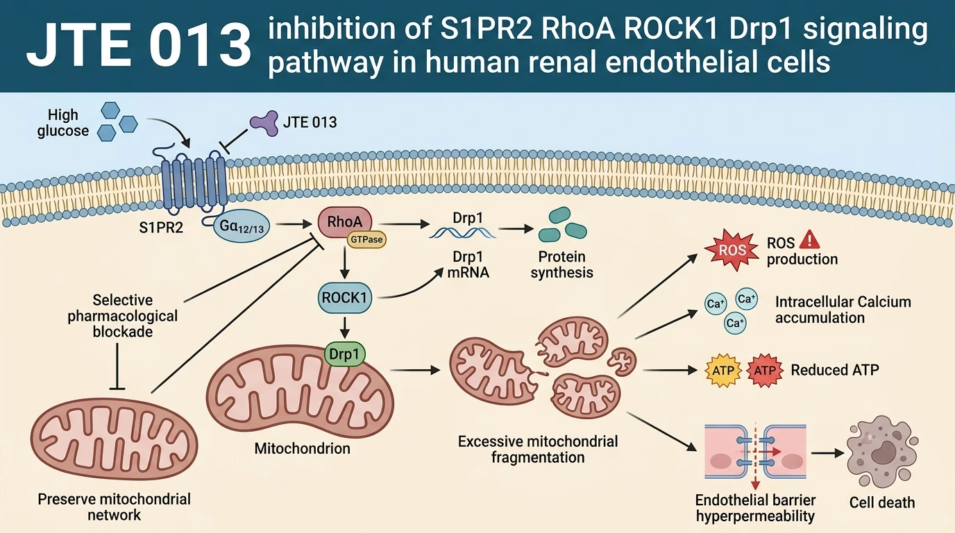
Sphingosine-1-phosphate receptor 2 (S1PR2), a membrane-bound G protein-coupled receptor, serves as a critical upstream mediator in transducing hyperglycemic stressors into intracellular structural damage. Upon stimulation, S1PR2 couples predominantly to G12/13 heterotrimeric G proteins, triggering the activation of the Small GTPase RhoA and its downstream effector kinase, Rho-associated coiled-coil containing protein kinase 1 (ROCK1). The RhoA/ROCK1 signaling cascade acts as an intracellular regulator of cytoskeletal assembly and organelle dynamics. Elevated glucose levels upregulate S1PR2 signaling, initiating sustained RhoA GTPase loading and ROCK1 kinase activation. This signal propagation upregulates expression of Dynamin-related protein 1 (Drp1), the primary GTPase responsible for mitochondrial outer membrane constriction and cleavage.
Experimental intervention using JTE 013, a selective small-molecule antagonist targeting S1PR2, demonstrates effective inhibition of this pathogenic pathway in cultured HRGECs. Pretreatment with JTE 013 directly blocks S1PR2 ligand binding and downstream G protein coupling. Assays performed in HG-exposed HRGECs confirm that JTE 013 application reduces transcript and protein levels of RhoA, ROCK1, and Drp1 to baseline control levels. By dampening the RhoA/ROCK1 signaling axis, the compound suppresses Drp1-mediated mitochondrial fission. Visual analysis of mitochondrial morphology shows restoration of elongated, reticulated mitochondrial networks in place of isolated, fragmented puncta.
Prevention of Drp1 upregulation by the JTE 013 S1P Receptor antagonist yields substantial secondary bioenergetic benefits. Mitigating mitochondrial fission limits electron transport chain electron leakage, significantly suppressing HG-induced intracellular ROS generation. Intracellular free calcium concentrations stabilize near non-diabetic control levels. Restoring mitochondrial structural integrity re-establishes normal oxidative phosphorylation, preventing the severe drop in cellular ATP levels observed in untreated HG cultures. Furthermore, endothelial cell functional assays indicate that S1PR2 receptor blockade rescues cell migration kinetics and decreases monolayer hyperpermeability, protecting HRGECs from glucose-induced apoptotic signaling cascades.
Direct biochemical comparison confirms that pharmacological ROCK1 inhibition mimics the cytoprotective profile of upstream S1PR2 blockade. When ROCK1 activity is suppressed directly, Drp1 expression declines in tandem, preventing HG-induced mitochondrial fragmentation and endothelial apoptosis. These concordant results validate the S1PR2/RhoA/ROCK1/Drp1 axis as a cohesive linear cascade regulating mitochondrial morphology in renal microvascular cells. In vitro findings establish S1PR2 receptor antagonism as an effective pharmacologic approach to preserve endothelial cell mitochondrial networks under high-glucose conditions, positioning this pathway for further evaluation in diabetic microvascular pathology models.
S1PR2 Antagonism Preserves Endothelial Mitochondrial Integrity
Copy code to add this image to your page
JTE-013 acts as a selective antagonist of sphingosine-1-phosphate receptor 2 (S1PR2). In human renal glomerular endothelial cells exposed to high glucose, S1PR2 activation increases expression of RhoA, ROCK1, and Drp1, causing excessive mitochondrial fission, ROS generation, calcium overload, ATP depletion, increased permeability, apoptosis, and impaired migration. JTE-013 blocks S1PR2, downregulating the RhoA/ROCK1/Drp1 pathway, preventing Drp1-mediated mitochondrial fragmentation, and restoring mitochondrial function and cellular viability.
S1PR2 Blockade Suppresses RhoA ROCK1 Drp1 Induction in Glomerular Endothelium
Exposure of human renal glomerular endothelial cells (HRGECs) to elevated extracellular glucose induces rapid transcriptional up-regulation of RhoA, ROCK1, and Drp1. This molecular cascade drives pathological mitochondrial fragmentation. In this cellular model, pretreatment with the selective JTE 013 S1P Receptor antagonist attenuates S1PR2-mediated signaling events. RhoA expression levels decreased substantially in drug-treated groups. Consequently, downstream ROCK1 activation and Drp1 accumulation dropped to physiological basal levels. Mitochondrial network morphology was preserved. Reticulated mitochondrial structures replaced isolated puncta under high-glucose culture conditions. These observations indicate that S1PR2 activity is required for glucose-induced Drp1 expression.
Mitochondrial Structural Preservation Restores Bioenergetic Output and Cell Migration
Uncontrolled mitochondrial fission triggers cellular bioenergetic crisis and endothelial barrier dysfunction. High-glucose challenged HRGECs exhibited increased intracellular calcium accumulation and decreased cellular ATP concentrations. Pretreatment with the JTE 013 S1P Receptor antagonist reversed these biochemical alterations. Intracellular reactive oxygen species generation declined markedly. Mitochondrial membrane potential stabilized near control values. At the functional level, treated endothelial cells exhibited increased migration capacity and reduced monolayer permeability. Endothelial apoptosis was suppressed. These pharmacodynamic outcomes demonstrate that targeting S1PR2 mitigates glucose-mediated endothelial dysfunction through structural and metabolic restoration.
Pharmacological ROCK1 Inhibition Replicates S1PR2 Antagonist Cytoprotection
To confirm the downstream mechanics of the S1PR2 axis, selective ROCK1 inhibitors were tested alongside receptor blockade. Direct inhibition of ROCK1 reduced Drp1 protein expression without altering upstream S1PR2 levels. High glucose-induced mitochondrial fragmentation was attenuated to an equivalent extent. Intracellular ROS accumulation and apoptotic rate were significantly lower in treated cells. These in vitro findings confirm that ROCK1 functions as the critical intermediate linking S1PR2 activation to Drp1-driven mitochondrial fission. The JTE 013 S1P Receptor antagonist acts upstream to suppress this destructive enzymatic cascade.