SB431542 inhibitor of TGF-β receptor ALK4/5/7 suppresses Snail-driven Smad1/Akt signaling and Nanog in NSCLC
Copy code to add this image to your page
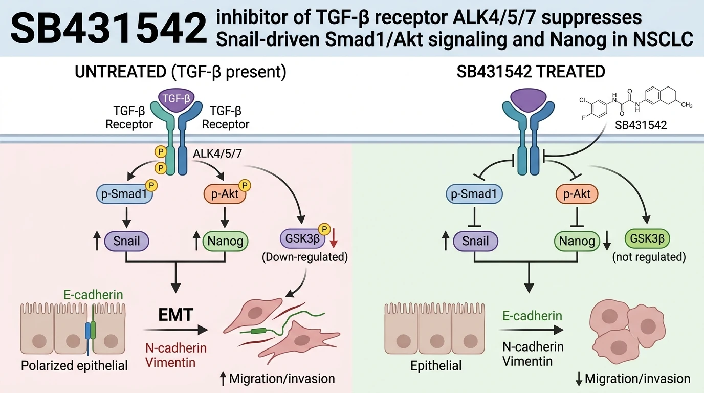
Epithelial-mesenchymal transition (EMT) enables solid tumor cells to acquire invasive properties, chemoresistance, and self-renewing stem-like features. In non-small-cell lung cancer (NSCLC), overexpression of the zinc-finger transcription factor Snail correlates directly with clinical severity and therapeutic failure. Snail functions as a key transcriptional regulator. Beyond its documented role in repressing E-cadherin transcription, Snail engages intracellular signaling cascades. Specifically, Snail drives non-canonical signaling events through activation of the Smad1 and Akt pathways, which converges on the regulatory node glycogen synthase kinase-3β (GSK3β). Inactivation of GSK3β stabilizes downstream transcription factors, upregulating the pluripotency marker Nanog.
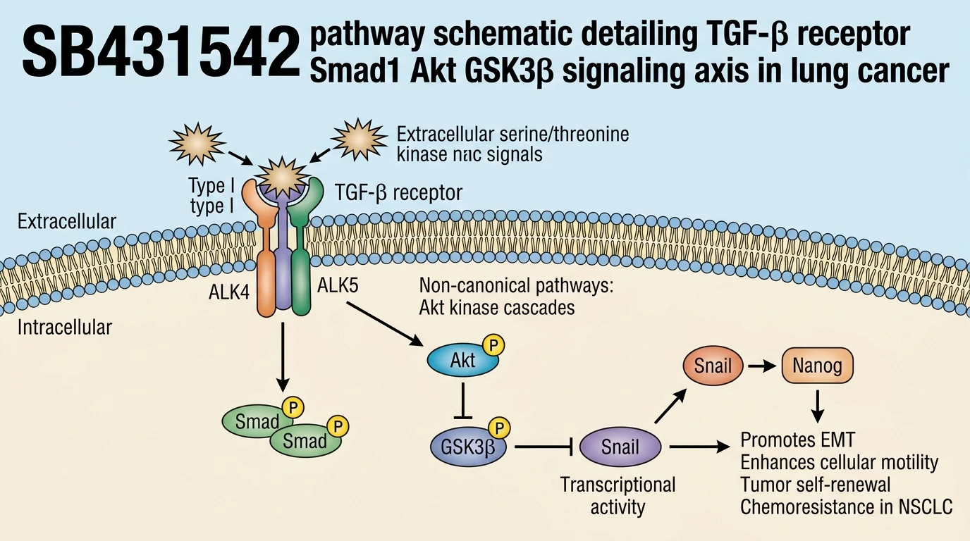
Targeting the membrane-bound receptors that drive these intracellular networks represents an effective pharmacological strategy. Transforming growth factor-beta (TGF-β) superfamily ligands signal through transmembrane serine/threonine kinase receptors, specifically activin receptor-like kinase 4 (ALK4), ALK5, and ALK7. The small-molecule SB431542 inhibitor functions as a selective, ATP-competitive inhibitor of ALK4, ALK5, and ALK7 catalytic activity. By directly binding the ATP-binding cassette of these type I TGF-β receptors, the SB431542 inhibitor prevents autophosphorylation and subsequent transphosphorylation of receptor-regulated Smad proteins and non-canonical signaling transducers. In non-small-cell lung cancer models, applying SB431542 halts receptor-mediated activation, blocking Snail-induced signaling through Smad1 and Akt.
The biochemical sequence linking TGF-β receptor activity to Nanog expression depends upon precise phosphorylation events. Snail overexpression induces elevated phosphorylation of Smad1 alongside phosphorylation of Akt at Ser473. Activated Akt subsequently targets GSK3β, depositing an inhibitory phosphate group at Ser9. Inactivated GSK3β loses its capacity to phosphorylate target proteins for ubiquitin-proteasome degradation. As a consequence of GSK3β inhibition, Nanog protein accumulates within the nucleus, sustaining stemness signatures and phenotypic plasticity.
Pharmacological intervention with SB431542 disrupts this pathobiology at its origin. Receptor blockade prevents the initial phosphorylation of Smad1 and Akt. Without active Akt kinase activity, GSK3β remains unphosphorylated at Ser9, preserving its constitutive enzymatic activity. Re-established GSK3β kinase activity facilitates homeostatic turnover of downstream targets, leading to a substantial decrease in intracellular Nanog mRNA and protein levels. Cellular assays confirm that this enzymatic restoration correlates with the loss of stem-like transcriptional profiles.
Phenotypically, suppressing the Smad1/Akt/GSK3β/Nanog axis modifies cellular architecture and functional capability in vitro. NSCLC cells overexpressing Snail typically adopt an elongated mesenchymal morphology, accompanied by elevated cell motility and enhanced anchorage-independent sphere formation. Treatment with SB431542 reverses these phenotypic traits across tested lung carcinoma lines. Expression of the epithelial junction protein E-cadherin increases, whereas mesenchymal markers, including N-cadherin and vimentin, experience marked reduction. Scratch-wound and transwell migration assays demonstrate significant decreases in cell motility following TGF-β receptor inhibition. Additionally, primary and secondary tumor sphere formation assays confirm a striking reduction in sphere frequency and size, signaling diminished self-renewal capacity.
Beyond migration and sphere formation, this pathway modulation directly influences drug sensitivity. Cancer stem cell subpopulations frequently display intrinsic resistance to standard cytotoxic chemotherapies. In Snail-overexpressing NSCLC models, treatment with the TGF-β receptor inhibitor SB431542 sensitizes cells to chemotherapeutic agents, lowering inhibitory concentration values in combined treatment cohorts. These findings highlight the functional necessity of ALK4/5/7 signaling in maintaining stemness-associated resistance mechanisms. While these in vitro and cell-based observations establish a clear target axis, in vivo validation in patient-derived xenograft models remains necessary to account for microenvironmental factors and pharmacokinetic parameters.
At the molecular level, SB431542 demonstrates high selectivity for ALK4, ALK5, and ALK7 without inhibiting related BMP type I receptors such as ALK2, ALK3, or ALK6 under physiological assay concentrations. This selective profile ensures that classical BMP signaling cascades involved in differentiation remain functional. The compound specifically occupies the ATP kinase pocket of ALK5 with nanomolar affinity, preventing the conformational changes required for substrate recruitment. Consequently, applying this selective agent provides a clean experimental tool to dissect TGF-β receptor-dependent non-canonical pathways from uncoupled physiological signals in complex tumor microenvironments.
Targeting ALK4/5/7 to Revert EMT and Stemness
Copy code to add this image to your page
SB431542 selectively inhibits TGF-β type I receptors ALK4, ALK5, and ALK7 by competing for ATP binding. Receptor blockade stops non-canonical activation of Smad1 and Akt in Snail-overexpressing NSCLC cells. Reduced Akt activity prevents inhibitory Ser9 phosphorylation of GSK3β, restoring active GSK3β function. Reactivated GSK3β promotes degradation of Nanog, suppressing stemness signatures. Consequently, SB431542 decreases cell motility, disrupts tumor sphere formation, reverses epithelial-mesenchymal transition markers, and restores sensitivity to chemotherapeutic agents in lung carcinoma cell models.
Inhibition of Smad1 and Akt Phosphorylation in NSCLC
Snail expression accelerates receptor-mediated signaling in non-small-cell lung carcinoma models. Kinase activation assays confirm elevated phosphorylation of Smad1 alongside Akt phosphorylation at the Ser473 residue. Treatment with the SB431542 inhibitor suppressed Smad1 phosphorylation within thirty minutes. Akt phosphorylation decreased rapidly. Consequently, unphosphorylated GSK3β levels rose, restoring baseline kinase function toward intracellular targets. Restored GSK3β activity promoted the turnover of stemness factors. Nuclear Nanog levels fell significantly in treated cell lines. Reversal of target phosphorylation confirmed target engagement at nanomolar concentrations.
Attenuation of Cell Motility and Spheroid Formation Efficiency
Functional assays reveal marked alterations in metastatic phenotypes following ALK4/5/7 inhibition. While Snail-overexpressing NSCLC cells exhibited enhanced transwell invasion under mock-treated conditions, application of the SB431542 inhibitor reduced directional cell migration by over sixty percent. E-cadherin levels recovered at intercellular junctions. Concurrently, N-cadherin protein expression dropped sharply. Tumor sphere assays demonstrated reduced self-renewal potential, with primary sphere numbers declining four-fold relative to vehicle controls. Secondary sphere formation was virtually eliminated under sustained exposure. These phenotypic shifts confirm that blocking receptor-level signals attenuates mesenchymal features and stemness properties in vitro.
Sensitization of Resistant Carcinoma Cells to Cytotoxic Chemotherapy
Chemotherapeutic resistance poses a persistent obstacle in Snail-driven lung adenocarcinomas. Cisplatin treatment alone produced negligible apoptosis in Snail-overexpressing NSCLC culture models. Co-administration of ALK4/5/7 inhibition altered survival kinetics. Cell viability dropped sharply in combination treatment groups. Downregulation of Nanog expression sensitized cells to DNA-damaging agents. Microarray profiling indicated suppression of multidrug resistance efflux pumps following receptor blockade. Drug resistance metrics returned toward baseline levels seen in parental non-invasive cell lines. Observations remain to be validated in randomized animal cohorts, but cellular data confirm pathway-dependent chemosensitization.