EHop-016 and EHop016 Suppress Rac1-Driven Glycolysis to Overcome Sorafenib Resistance
Copy code to add this image to your page
Primary therapy for advanced hepatocellular carcinoma (HCC) frequently fails due to acquired or intrinsic resistance to sorafenib, a multikinase inhibitor targeting RAF, VEGFR, and PDGFR. Aberrant metabolic adaptation—specifically enhanced aerobic glycolysis, commonly termed the Warburg effect—serves as a predominant mechanism fueling cell survival under chemotherapeutic pressure. Emerging molecular profiles identify Ras-associated C3 botulinum toxin subunit 1 (Rac1), a small Rho family GTPase, as an indispensable driver of oncogenic growth and metabolic plasticity in HCC tissues. Clinical observations correlate elevated Rac1 overexpression with reduced overall patient survival and persistent sorafenib refractory phenotypes.
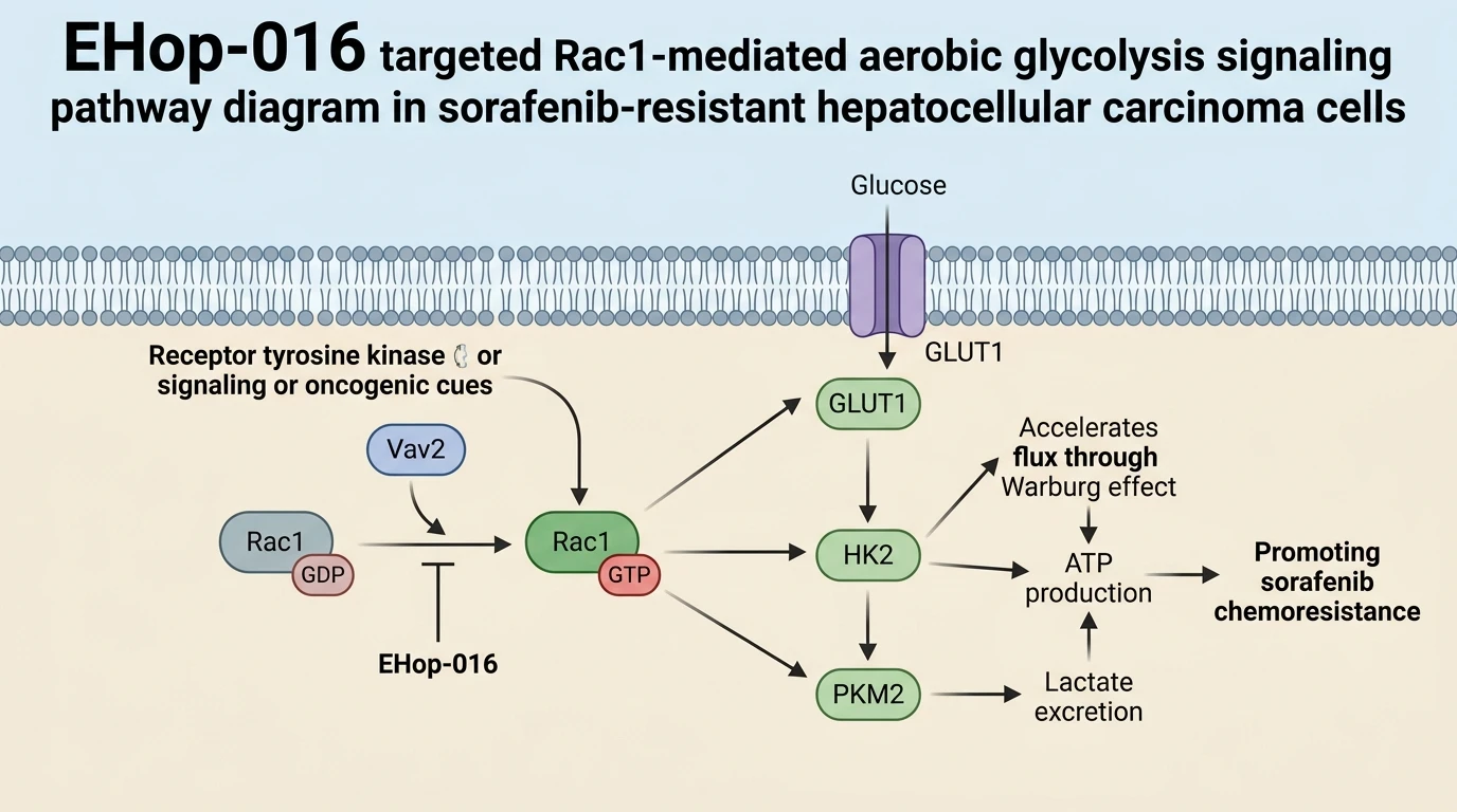
At the biochemical level, Rac1 functions as a molecular switch transitioning between an inactive GDP-bound form and an active GTP-bound state. Guanine nucleotide exchange factors (GEFs) such as Vav2 catalyze GTP loading, enabling Rac1 to engage downstream effectors that regulate actin cytoskeletal dynamics, cell cycle progression, and transcriptional programs governing energy metabolism. In sorafenib-resistant HCC lines, constitutively active Rac1 sustains high glycolytic flux by driving the membrane translocation of glucose transporter 1 (GLUT1) and transcriptionally activating key rate-limiting enzymes such as hexokinase 2 (HK2) and pyruvate kinase M2 (PKM2). This shift accelerates intracellular glucose uptake, elevates ATP production through substrate-level phosphorylation, and leads to excessive lactate export. Consequently, tumor cells bypass sorafenib-induced metabolic stress, preserving mitochondrial integrity and avoiding apoptotic signaling cascades.
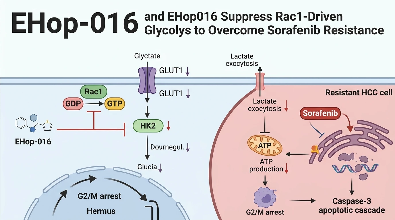
Direct pharmacologic target engagement of Rac1 presents a targeted approach to disrupt this bioenergetic adaptation. EHop-016 acts as a small-molecule inhibitor designed to target Rac1 activation by blocking its interaction with specific GEFs. In cell-free binding assays, EHop-016 selectively binds the Rac1 protein with IC50 values in the low micromolar range (approximately 1.1 μM), inhibiting GEF-catalyzed nucleotide exchange without altering closely related GTPases such as Cdc42 or RhoA at equivalent dosages. In cultured HCC cell models displaying sorafenib tolerance, administration of EHop-016 or its derivative formulation EHop016 suppresses Rac1-GTP loading within hours of treatment. The resulting blockade of downstream signaling impairs p21-activated kinase 1 (PAK1) activation, directly compromising the cell glycolytic infrastructure.
Experimental evaluation demonstrates that Rac1 inhibition via EHop-016 dismantles the bioenergetic buffer of HCC cells. In sorafenib-resistant cell lines under tested assay conditions, combined treatment with EHop-016 and sorafenib yields a marked decrease in glucose consumption and intracellular lactate production relative to sorafenib monotherapy. ATP quantification assays show a concurrent depletion of cellular energy reserves under these co-treatment regimes. This metabolic deprivation destabilizes the mitochondrial membrane potential, promoting cytochrome c release and triggering activation of caspase-9 and caspase-3. Flow cytometric analysis further verifies that EHop-016 administration combined with sorafenib induces pronounced G2/M phase cell cycle arrest, arresting proliferative expansion and triggering apoptotic execution pathways.
In vivo evaluation utilizing murine xenograft models bearing sorafenib-resistant HCC tumors supports these cellular observations. Systemic administration of EHop-016 in combination with sorafenib results in significant tumor volume reduction compared to single-agent controls. Immunohistochemical and Western blot analyses of harvested tumor tissue reveal attenuated expression of Rac1-GTP, decreased HK2 levels, and elevated markers of apoptosis such as cleaved PARP. Furthermore, mice tolerated the combined pharmacologic regimen without displaying overt systemic toxicity or weight loss within the experimental timeframe. These findings indicate that targeting Rac1 with EHop-016 provides a viable strategy to suppress aerobic glycolysis and overcome sorafenib chemoresistance in hepatocellular carcinoma, offering a rationale for further translational investigation.
Therapeutic Evaluation and Pharmacological Characterization
Copy code to add this image to your page
EHop-016 directly inhibits Rac1 activation by blocking guanine nucleotide exchange factor binding, thereby downregulating aerobic glycolysis in hepatocellular carcinoma cells. Suppressing Rac1-GTP levels lowers glucose uptake, reduces lactate production, and depletes cellular ATP reserves. When combined with sorafenib, EHop016 disrupts bioenergetic compensation, induces G2/M cell cycle arrest, and triggers intrinsic caspase-dependent apoptosis. In vivo xenograft studies confirm that pharmacologic Rac1 inhibition restores sorafenib sensitivity and suppresses tumor growth, validating Rac1 targeted therapy to overcome sorafenib resistance.
Attenuation of Rac1-GTP Loading Suppresses Glycolytic Enzymes in Vitro
Target engagement assays confirm that Rac1 inhibition impairs glycolytic machinery in sorafenib-resistant HCC cells. Treatment with EHop-016 reduced active Rac1-GTP levels in a concentration-dependent manner without affecting total Rac1 protein expression. Downstream phosphorylation of PAK1 ceased within two hours of exposure. Under these in vitro culture conditions, GLUT1 membrane expression dropped significantly. Intracellular glucose uptake fell by over 45% relative to vehicle-treated controls. Consequently, key rate-limiting glycolytic enzymes—including HK2 and PKM2—showed marked transcriptional and translational downregulation. Lactate excretion into the culture media diminished concurrently. Cellular ATP levels declined by more than half following 24 hours of sustained exposure. These empirical metrics demonstrate that targeting Rac1 directly disrupts the bioenergetic infrastructure sustaining tumor cell survival.
Combination with Sorafenib Induces Apoptosis and Arrests Cell Cycle
Co-administration of sorafenib and EHop016 restored chemotherapeutic sensitivity in refractory cell lines. Sorafenib alone failed to trigger substantial cell death in resistant models at standard doses. The combination regime altered cell fate. Cellular viability dropped sharply following co-treatment. Flow cytometry demonstrated a clear enrichment of cells in the G2/M phase, indicating mitotic arrest. Immunoblotting revealed elevated cleavage of initiator caspase-9 and executioner caspase-3 within 12 hours. Annexin V staining confirmed a three-fold increase in early and late apoptotic populations. This synergy indicates that metabolic suppression via Rac1 blockade sensitizes resistant cells to multikinase inhibitor-induced apoptosis.
Murine Xenograft Suppression Confirms In Vivo Chemosensitization
In vivo efficacy was validated in a immunodeficient mouse xenograft model using sorafenib-resistant HCC cell lines. Vehicle control groups and sorafenib monotherapy cohorts exhibited rapid tumor burden progression. Combined treatment with EHop-016 and sorafenib attenuated tumor growth significantly over the 21-day treatment window. Final tumor weights in the combination cohort were reduced by 62% relative to sorafenib single-agent controls. Western blot analysis of xenograft tissue lysates verified marked reductions in Rac1-GTP levels and HK2 expression. Histological examination showed increased cleaved PARP staining in tumor sections from combination-treated mice. Animals maintained normal body weight and displayed no overt clinical signs of systemic toxicity. These observations remain to be validated in clinical cohorts, but they establish a firm rationale for targeting Rac1 to overcome sorafenib resistance.