Etomoxir sodium salt suppresses CPT1-mediated fatty acid oxidation to rescue Mstn-deficient HeLa apoptosis
Copy code to add this image to your page
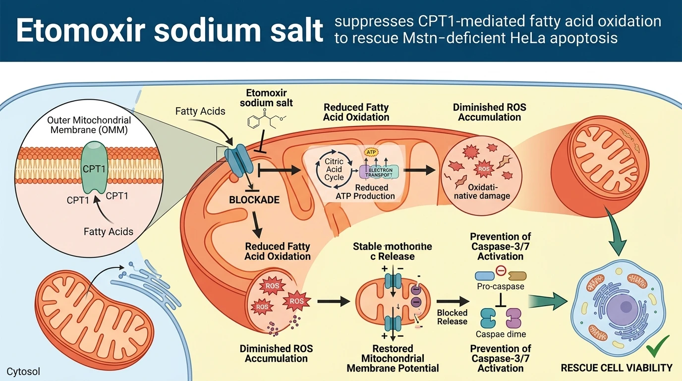
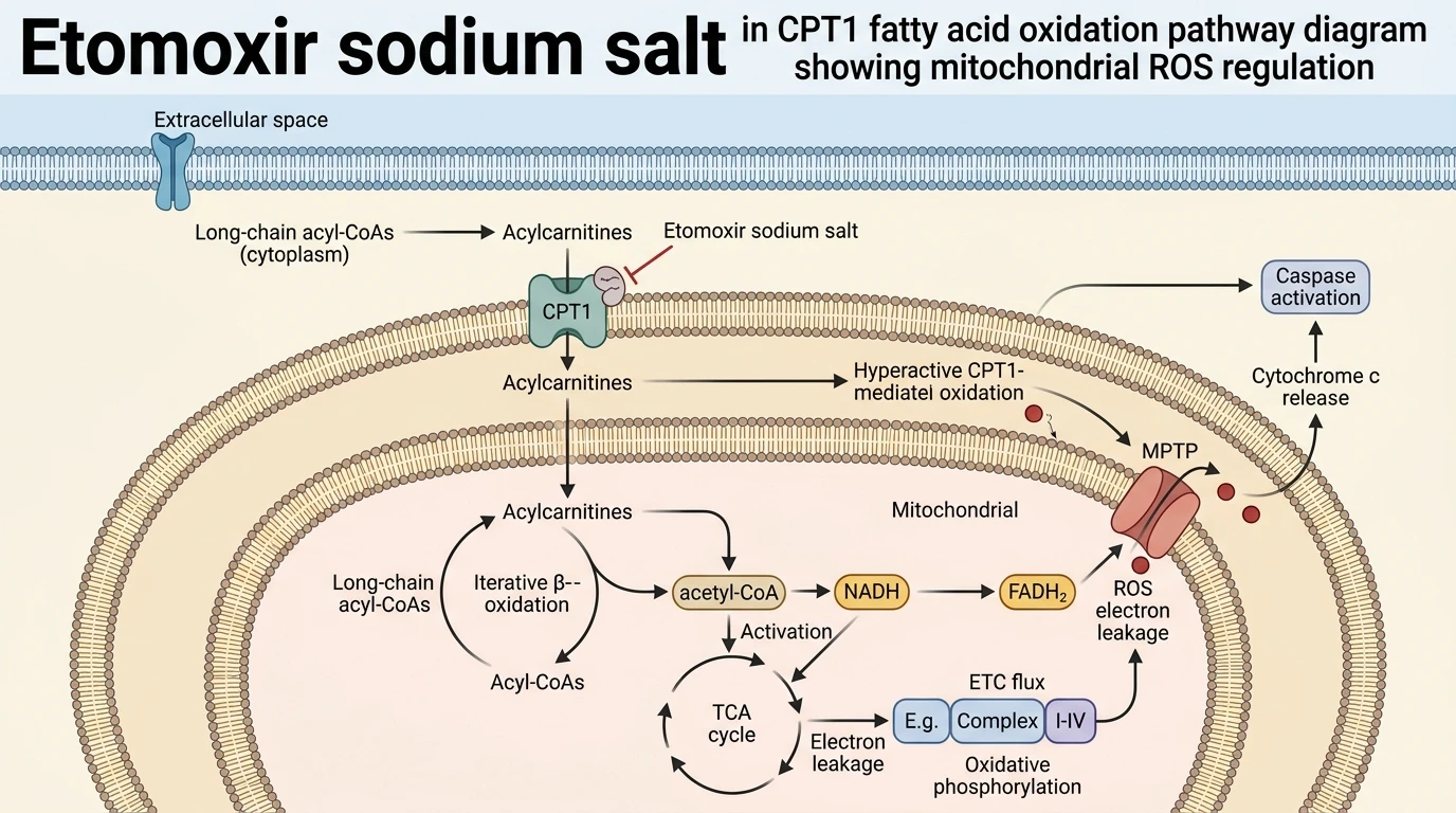
Genetic disruption of Myostatin (Mstn) in human cervical adenocarcinoma (HeLa) cells alters metabolic programming, shifting energetic dependence toward fatty acid oxidation (FAO). Under tested in vitro assay conditions, Mstn knockout markedly upregulates key transcripts governing lipid catabolism, prominently inducing carnitine palmitoyltransferase 1 (CPT1). CPT1 serves as the rate-limiting translocase responsible for shuttling long-chain fatty acyl-CoA esters into the mitochondrial matrix. Elevated expression of this outer mitochondrial membrane enzyme accelerates fatty acid oxidation flux beyond homeostatic thresholds. Accelerated lipid oxidation directly intensifies electron transport chain activity, generating excessive mitochondrial reactive oxygen species (ROS). Intracellular ROS overload subsequently destabilizes organellar homeostasis in Mstn-deficient HeLa cells. Mitochondrial membrane potential decreases rapidly following the oxidative surge, accelerating outer membrane permeabilization. Cytosolic accumulation of cytochrome c occurs within hours of peak ROS generation. Released cytochrome c binds apoptotic protease activating factor-1 to assemble the apoptosome, initiating downstream executioner cascades; fluorometric assays confirm marked elevation of cleaved caspase-3 and caspase-7 activity. Small-molecule intervention using Etomoxir sodium salt directly disrupts this pathological metabolic cascade. As a selective inhibitor of CPT1, Etomoxir binds the enzyme to prevent long-chain acylcarnitine formation. Pharmacological suppression of CPT1 restricts fatty acyl entry into the mitochondrial matrix, rapidly reducing matrix beta-oxidation rates and downstream electron leakage. At micromolar concentrations in cell culture models, CPT1 blockade attenuates ROS generation, preserves mitochondrial membrane potential, prevents cytosolic cytochrome c release, and suppresses caspase cleavage. These empirical observations establish that CPT1-mediated fatty acid oxidation drives ROS-induced intrinsic apoptosis in Mstn-deficient cervical cancer cells.
Pharmacological CPT1 Inhibition and Apoptotic Rescue Mechanics
Copy code to add this image to your page
Etomoxir sodium salt acts as a selective inhibitor of carnitine palmitoyltransferase 1 (CPT1), the rate-limiting enzyme in mitochondrial fatty acid oxidation. In Mstn-deficient HeLa cells, CPT1 expression and fatty acid oxidation are aberrantly elevated, generating excessive reactive oxygen species (ROS) that trigger mitochondrial depolarization, cytochrome c release, and caspase-3/7-mediated apoptosis. By inhibiting CPT1, Etomoxir reduces fatty acid oxidation rates, attenuates mitochondrial ROS accumulation, preserves membrane integrity, and rescues Mstn KO-induced apoptosis.
CPT1 Upregulation Drives Intracellular ROS Generation in Mstn KO HeLa
Myostatin deficiency fundamentally rewires cellular metabolism in HeLa models. Transcriptional profiling reveals robust upregulation of CPT1 following gene knockout. Enhanced enzyme density accelerates fatty acid translocation across the mitochondrial membrane; this metabolic shift increases respiratory chain flux and yields cytotoxic ROS levels. In vitro measurements confirm mitochondrial membrane depolarization. Cytochrome c release follows. Subsequently, executioner caspase-3 and caspase-7 undergo cleavage to execute intrinsic apoptosis.
Targeted CPT1 Inhibition Suppresses ROS and Restores Viability
Pharmacological administration of Etomoxir selectively blocks CPT1 enzymatic activity. Substrate availability for beta-oxidation drops immediately upon target engagement. As lipid catabolism declines, mitochondrial ROS production returns to baseline levels—preserving membrane potential and halting cytochrome c efflux into the cytosol. Apoptotic markers drop significantly. Cellular viability rises in a dose-dependent manner. These observations confirm that CPT1-driven lipid oxidation acts as the primary mechanistic trigger of cell death following Mstn ablation in this cell line.