Bemcentinib (R428) and Axl Kinase Inhibition in Gas6-SOCS3 Pulmonary Inflammatory Signaling
Copy code to add this image to your page
Ischemia-reperfusion-induced acute lung injury (IR-ALI) represents a profound clinical challenge characterized by severe alveolar-capillary membrane disruption, microvascular leakage, pulmonary edema, and intense parenchymal inflammation. During the reoxygenation phase following an ischemic episode, excessive reactive oxygen species production and massive release of pro-inflammatory cytokines trigger extensive damage to alveolar epithelial and endothelial barriers. Among the cell-surface receptors regulating innate immune responses and cell survival during hyperinflammatory states, the TAM family of receptor tyrosine kinases—comprising Tyro3, Axl, and MerTK—plays an important counter-regulatory role. Within this receptor family, Axl receptor tyrosine kinase (Axl) has emerged as a distinct regulator of tissue homeostasis and inflammatory resolution, binding with high affinity to its endogenous ligand, Growth Arrest-Specific 6 (Gas6). Insights into small-molecule modulation of kinase pathways have enhanced our understanding of these target interactions, as reflected in recent developments with small-molecule kinase inhibitors.
When Gas6 engages the extracellular ligand-binding domain of Axl, it induces receptor homodimerization and autophosphorylation of intracellular tyrosine residues (p-Axl). This phosphorylation event initiates downstream signaling cascades that modulate inflammatory gene expression and cellular survival pathways. In isolated rat lung models subjected to warm ischemia-reperfusion, ex vivo perfusion with recombinant Gas6 attenuated key structural and biochemical manifestations of acute lung injury. Specifically, Gas6 administration reduced gross lung weight gain and microvascular permeability metrics, while significantly lowering pulmonary edema index values. Histopathological examination revealed preserved alveolar capillary wall architecture with reduced neutrophilic infiltration and alveolar hemorrhage compared to untreated ischemic controls. Concurrently, levels of pro-inflammatory cytokines—including Tumor Necrosis Factor-alpha (TNF-α), Interleukin-1 beta (IL-1β), and Interleukin-6 (IL-6)—were substantially lower in bronchoalveolar lavage fluid following Gas6 administration.
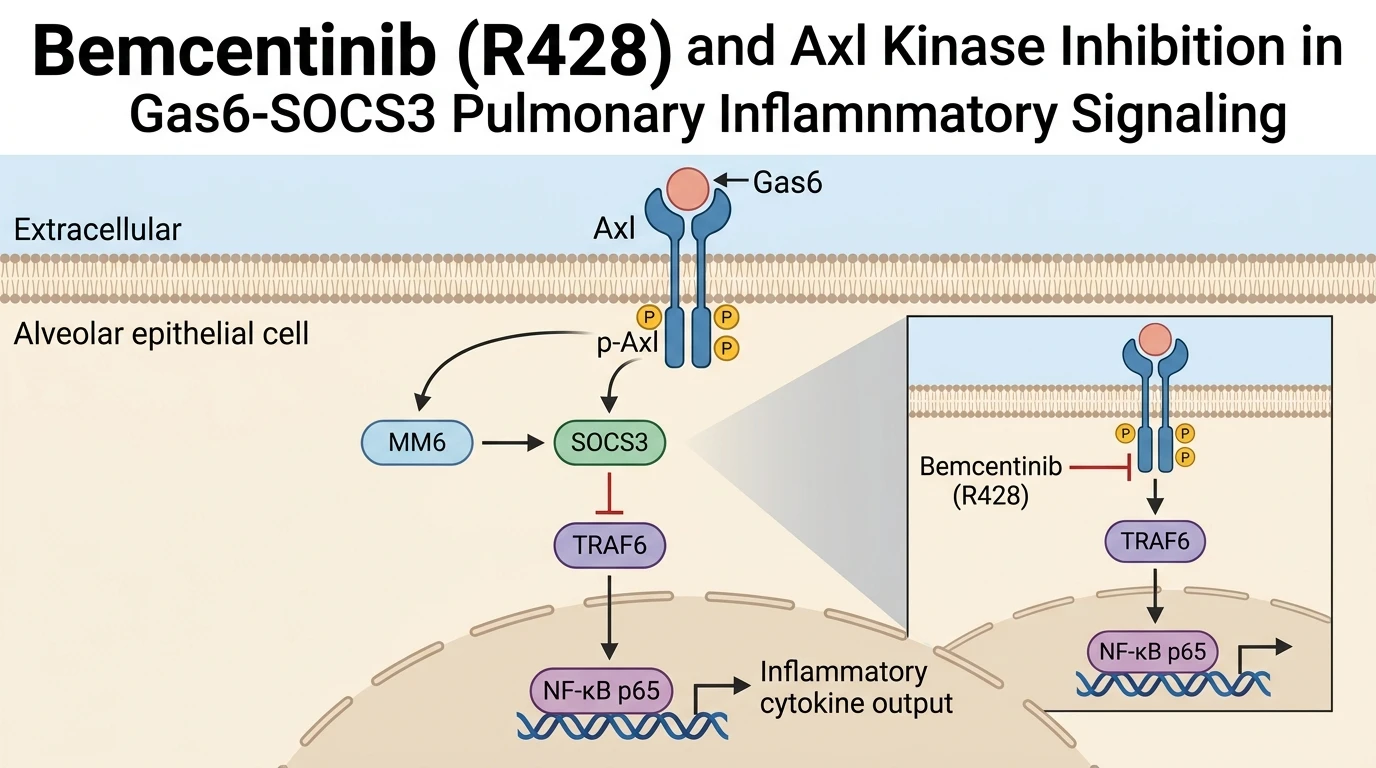
At the cellular level, investigation in MLE-12 mouse lung alveolar epithelial cells subjected to hypoxia-reoxygenation (HR) provided deeper insight into the intracellular mechanics downstream of p-Axl. Hypoxia followed by reoxygenation severely depleted endogenous p-Axl and Suppressor of Cytokine Signaling 3 (SOCS3) expression. SOCS3 is an established feedback inhibitor of cytokine signaling that downregulates signal transduction through intracellular adaptor proteins. The decline in SOCS3 under HR conditions correlated directly with elevated expression of Tumor Necrosis Factor Receptor-Associated Factor 6 (TRAF6) and enhanced nuclear translocation of the Nuclear Factor Kappa B (NF-κB) p65 subunit. Active nuclear NF-κB p65 acts as a master transcriptional activator of pro-inflammatory mediators, driving the hyperinflammatory cascade observed in pulmonary injury.
Exogenous application of Gas6 to HR-treated MLE-12 epithelial cells restored p-Axl levels in a concentration-dependent manner, directly accompanied by robust upregulation of SOCS3 protein expression. Elevated SOCS3 engaged TRAF6, inhibiting its ubiquitination signaling activity and suppressing the translocation of NF-κB p65 into the nucleus. This mechanistic axis establishes that Gas6/Axl signaling limits alveolar inflammation primarily by enforcing SOCS3-dependent suppression of the TRAF6/NF-κB pathway.
To rigorously evaluate whether Axl enzymatic activity is required for SOCS3 induction and anti-inflammatory protection, pharmacologic target antagonism was performed using Bemcentinib (R428). Pretreatment of MLE-12 cells with the selective Axl inhibitor R428 completely abolished Gas6-induced p-Axl phosphorylation. In the presence of R428, Gas6 failed to upregulate SOCS3 expression, leading to full restoration of TRAF6 accumulation and nuclear p65 localization. Similarly, in ex vivo rat lung preparations, co-administration of R428 negated the protective effects of Gas6 on microvascular permeability and cytokine release. These pharmacological perturbation experiments with R428 confirm that catalytic activity of Axl is indispensable for initiating the protective SOCS3 signal transduction cascade in pulmonary models of ischemia-reperfusion injury.
Pharmacological Characterization of Axl Kinase in Ischemic Alveolar Injury
Copy code to add this image to your page
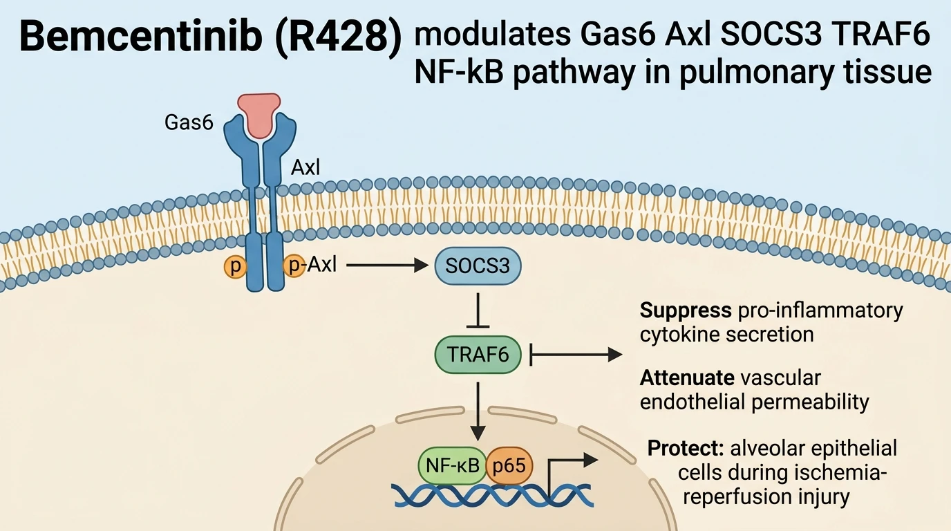
Gas6 activates Axl receptor tyrosine kinase phosphorylation (p-Axl), which drives expression of Suppressor of Cytokine Signaling 3 (SOCS3). Increased SOCS3 inhibits TRAF6, suppressing NF-κB p65 nuclear translocation and downregulating pro-inflammatory cytokine production in alveolar epithelial cells. In isolated rat lungs and MLE-12 cells under hypoxia-reoxygenation, this axis reduces edema, vascular permeability, and inflammation. Selective inhibition of Axl by Bemcentinib (R428) blocks SOCS3 upregulation, fully reversing the anti-inflammatory and tissue-protective effects of Gas6 during ischemia-reperfusion injury.
Gas6 Attenuates Microvascular Permeability in Isolated Rat Lungs
Ex vivo rat lung models provide a controlled biomechanical system to evaluate pulmonary ischemia-reperfusion injury. Under isolated perfused conditions, reperfusion following ischemia induced marked microvascular leakage, lung edema, and histological tissue degradation. Exogenous administration of Gas6 significantly mitigated these pathological parameters. Microvascular permeability markers returned toward baseline levels. Cytokine accumulation in the bronchoalveolar fluid decreased markedly. Mechanistically, this protection depended upon intact Axl phosphorylation. Phosphorylation of Axl activated downstream signaling pathways that preserve endothelial tight junctions and alveolar-capillary barrier integrity. These empirical observations establish Gas6 as a functional suppressor of ischemic vascular compromise.
Axl Activation Restores SOCS3 and Represses Nuclear NF-κB p65
In vitro hypoxia-reoxygenation (HR) assays in MLE-12 alveolar epithelial cells clarified the underlying intracellular cascade. Hypoxic stress suppressed baseline p-Axl and SOCS3 protein levels. Consequently, TRAF6 expression increased, accompanied by rapid nuclear translocation of the NF-κB p65 subunit. Recombinant Gas6 treatment fully restored p-Axl and SOCS3 expression in HR-exposed cells. Elevated SOCS3 directly suppressed TRAF6 activity. Nuclear p65 levels dropped significantly following treatment. To confirm target specificity, cells were pretreated with the selective small-molecule inhibitor R428. Inhibition of Axl phosphorylation by R428 entirely blocked SOCS3 upregulation. As a result, TRAF6 repression was abolished and NF-κB p65 nuclear translocation resumed. This pharmacological reversal by R428 proves that SOCS3 induction requires active Axl catalytic domain signaling.