RepSox (E-616452) Attenuates Cancer-Induced Bone Pain via ALK5 Smad3 Signaling Inhibition
Copy code to add this image to your page
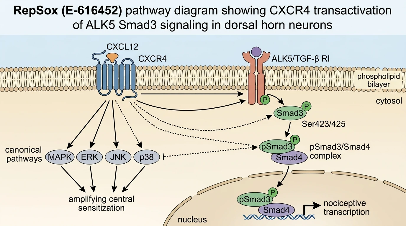
Cancer-induced bone pain (CIBP) presents a severe clinical challenge characterized by deep aching pain, mechanical allodynia, and thermal hyperalgesia. Pathophysiologically, tumor cell invasion into the bone marrow stroma triggers localized tissue acidosis, nerve sprouting, and sustained release of inflammatory mediators. These peripheral signals converge on the dorsal root ganglion and project directly to the superficial layers (laminae I–II) of the spinal dorsal horn. At the central synaptic junction, persistent chemokine receptor 4 (CXCR4) signaling induces neurochemical alterations in secondary nociceptive neurons and surrounding glial elements. Recent pharmacological evaluations indicate that spinal CXCR4 transactivates the activin receptor-like kinase 5 (ALK5), also recognized as transforming growth factor-beta type I receptor (TGFβRI). This transactivation promotes downstream phosphorylation of Smad3 (p-Smad3), establishing a central sensitization loop that maintains chronic pain phenotypes.
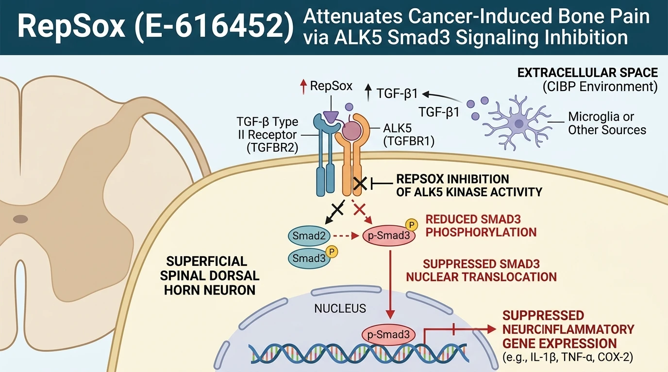
To selectively target this aberrant receptor cross-talk, researchers utilize small-molecule inhibitors of TGFβRI kinase activity. RepSox (E-616452) functions as an ATP-competitive, highly selective inhibitor of ALK5, displaying marked selectivity over closely related type I receptors such as ALK4 and ALK7 under in vitro kinase profiling assays. Mechanistically, RepSox binds the ATP-binding pocket of the ALK5 kinase domain, preventing autophosphorylation and subsequent substrate recruitment. In preclinical rodent models of tumor-induced bone destruction, intrathecal delivery of RepSox blocks the catalytic phosphorylation of Smad3 at Ser423/425 residues. Consequently, cytosolic p-Smad3 levels drop rapidly, impairing its association with Smad4 and halting nuclear translocation of the transcriptional complex.
In spinal cord nerve cells, ALK5 signaling operates through both canonical and non-canonical effector cascades. Canonical signaling relies directly on Smad3 transcription factor activation to upregulate pro-nociceptive target genes, including inflammatory cytokines and ion channel subunits. Non-canonical pathways branch into mitogen-activated protein kinase (MAPK) cascades, such as p38 MAPK and extracellular signal-regulated kinase (ERK), which modulate acute synaptic transmission. Within the superficial spinal dorsal horn of CIBP rats, both signaling axes display pronounced upregulation following tumor implantation. Experimental intrathecal administration of RepSox directly attenuates p-Smad3 accumulation across both canonical and non-canonical signaling nodes in these neural populations. This dual inhibitory mechanism attenuates nociceptive processing at the spinal level without compromising basal motor function.
Behavioral assessment in rat CIBP models demonstrates that central ALK5 blockade translates into significant relief from evoked-pain behaviors. Tumor-bearing animals subjected to intrathecal RepSox exhibit an attenuation of thermal hyperalgesia and mechanical allodynia, returning nociceptive withdrawal thresholds toward baseline levels. Mechanistic parsing suggests that CXCR4-driven transactivation of ALK5 requires membrane proximity and intracellular phosphorylation loops, which are dismantled upon small-molecule ALK5 inhibition. Importantly, these pharmacodynamic effects appear concentration-dependent within the tested intrathecal dose range. By uncoupling spinal chemokine receptor activity from downstream transcriptional remodeling, ALK5 inhibition offers a distinct pharmacological tactic for interrupting central sensitization in CIBP.
Pharmacological Evaluation of Spinal ALK5 Kinase Inhibition
Copy code to add this image to your page
RepSox acts as a potent ATP-competitive inhibitor of ALK5 (TGF-beta type I receptor kinase). By blocking ALK5 catalytic activity, RepSox prevents the transactivation-dependent phosphorylation of downstream Smad3 (p-Smad3) induced by spinal CXCR4 engagement. In rat models of cancer-induced bone pain, intrathecal RepSox suppresses both canonical Smad3 translocational signaling and non-canonical neuroinflammatory pathways in the spinal dorsal horn. This dual inhibition reduces central nociceptive sensitization, offering a novel mechanistic strategy for managing intractable bone pain.
Downregulation of Spinal Dorsal Horn p-Smad3 Expression
Immunohistochemical and immunoblotting analyses of spinal dorsal horn tissue reveal pronounced spatial upregulation of CXCR4, ALK5, and p-Smad3 following tumor cell inoculation. Expression localized predominantly to laminae I and II of the spinal cord dorsal horn. Intrathecal injection of RepSox markedly suppressed Ser423/425 phosphorylation on Smad3 without altering total ALK5 protein abundance. Western blot quantifications confirmed dose-dependent suppression of cytosolic and nuclear p-Smad3 fractions. Smad3 signaling ceased within hours post-administration. Crucially, non-canonical phosphorylation of p38 MAPK and ERK in spinal neuronal lysates also decreased following ALK5 inhibition. These biochemical shifts suggest that target engagement in the central nervous system limits broader neuroinflammatory gene networks.
Reversal of Nociceptive Sensitization in Rat Models
Sensory testing protocols established baseline paw withdrawal mechanical thresholds (PWMT) and paw withdrawal thermal latencies (PWTL) before and after intrathecal dosing regimes. In rat CIBP models, vehicle-treated animals displayed persistent mechanical allodynia and thermal hyperalgesia throughout the observation window. Administration of RepSox significantly elevated both PWMT and PWTL metrics relative to controls. Paw withdrawal thresholds rose within thirty minutes of intrathecal delivery. Therapeutic efficacy persisted across multiple post-injection intervals before returning toward baseline values. Motor coordination assays, including rotarod performance tests, confirmed no motor deficits at effective therapeutic doses. These functional metrics demonstrate that target engagement specifically targets nociceptive pathways.
CXCR4 Transactivation Dynamics and Receptor Crosstalk
The molecular link between chemokine sensing and TGF-beta receptor activation represents a critical node in spinal sensitization cascades. Stimulation of spinal CXCR4 by CXCL12 triggers rapid intracellular transactivation of ALK5, driving downstream Smad3 phosphorylation independently of canonical TGF-beta ligand binding. Co-immunoprecipitation experiments indicate physical interaction between CXCR4 and ALK5 complexes in spinal membrane fractions. Treatment with RepSox disrupted this receptor cross-talk by locking ALK5 in an inactive conformation. Downstream signaling events failed to materialize. Consequently, spinal neurons maintained baseline electrophysiological thresholds. These observations highlight receptor transactivation as a primary therapeutic vulnerability in cancer-induced pain states.