Celecoxib (SC-58635) COX inhibitor attenuates liver fibrosis via COX-2 PGE2 VEGF suppression
Copy code to add this image to your page
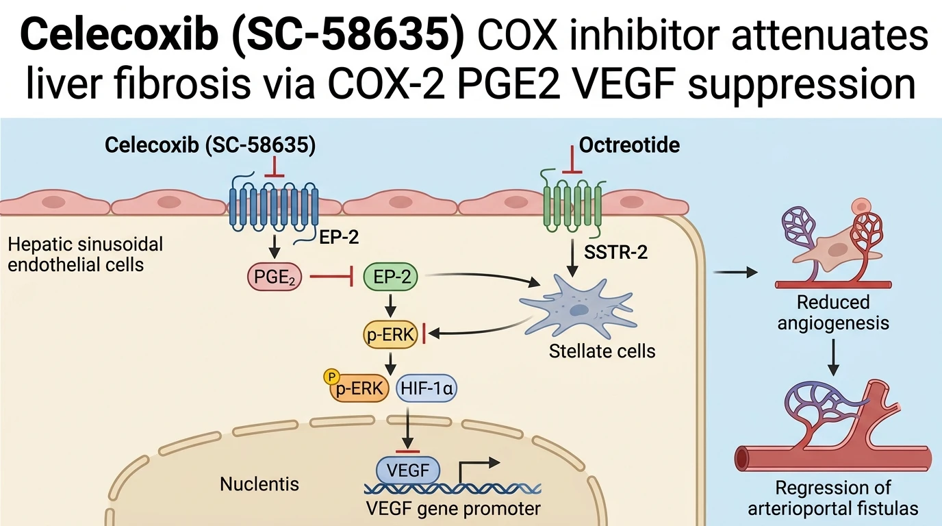
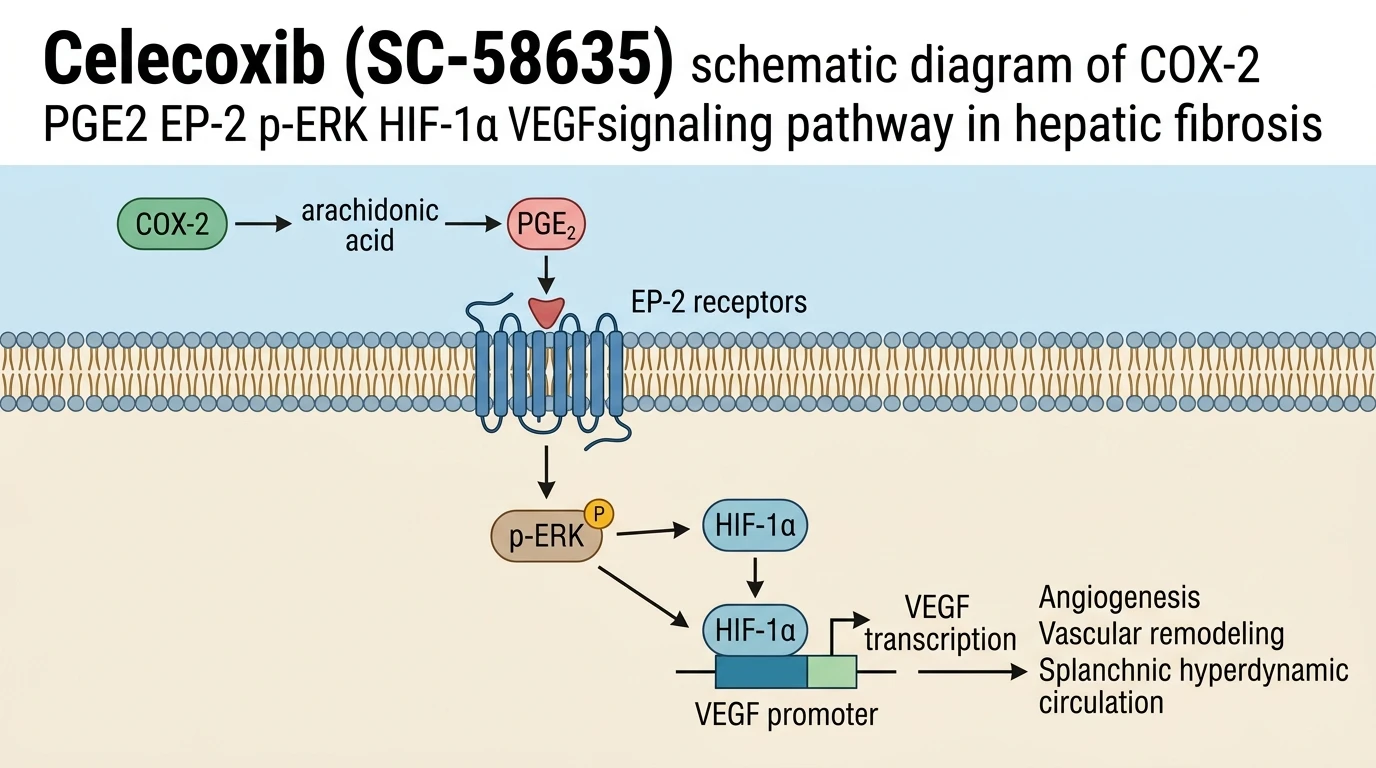
Cyclooxygenase-2 (COX-2) functions as an inducible rate-limiting enzyme responsible for catalyzing the conversion of arachidonic acid into prostaglandin H2, the precursor for prostaglandin E2 (PGE2). In chronic liver injury, such as thioacetamide (TAA)-induced hepatic cirrhosis, COX-2 expression increases markedly within non-parenchymal liver cells, including activated hepatic stellate cells, sinusoidal endothelial cells, and resident macrophages. Elevated local PGE2 concentrations signal through E-prostanoid receptor 2 (EP-2), activating intracellular G-protein cascades that stimulate downstream mitogen-activated protein kinase pathways. Phosphorylated extracellular signal-regulated kinase (p-ERK) subsequently stabilizes hypoxia-inducible factor-1α (HIF-1α), suppressing its ubiquitin-mediated proteasomal degradation and facilitating nuclear translocation. Inside the nucleus, HIF-1α heterodimerizes with HIF-1β and binds specific hypoxia response elements on the vascular endothelial growth factor (VEGF) promoter region, triggering transcription of pro-angiogenic factors. This pathway drives pathological intrahepatic angiogenesis, splanchnic hyperdynamic circulation, and structural remodeling that elevates portal venous pressure.
Pharmacological targeting of this axis using the selective Celecoxib (SC-58635) COX inhibitor provides a distinct biochemical mechanism for modulating pathological vessel formation without non-selective systemic cytotoxicity. In TAA-treated rat models of established liver cirrhosis, administrative regimens incorporating Celecoxib (SC-58635) reduced intrahepatic vascular resistance and splanchnic hyperperfusion. The molecule selectively binds the hydrophilic side-pocket of COX-2 formed by a single amino acid substitution (Val523 in COX-2 versus Ile523 in COX-1), effectively blocking substrate access to the active site. Inhibition of COX-2 enzymatic conversion reduces intrahepatic PGE2 levels, directly diminishing downstream EP-2 receptor occupation. As a consequence, cellular p-ERK accumulation declines, breaking the intracellular signaling loop required to maintain elevated HIF-1α expression under local tissue hypoxia.
Combinatorial therapeutic strategies targeting parallel anti-angiogenic nodes exhibit pronounced pharmacodynamic synergy in preclinical models. When co-administered with octreotide—a synthetic octapeptide somatostatin analogue acting predominantly via somatostatin receptor subtype 2 (SSTR-2)—the combined regimen demonstrates superior efficacy compared to either monotherapy alone. In rodent models of severe portal hypertension, dual treatment yielded statistically significant reductions in mean portal pressure and portosystemic shunting percentages. Splanchnic hemodynamics displayed marked attenuation, characterized by reduced superior mesenteric artery blood flow and normalized cardiac output parameters. Histological reconstructions and micro-computed tomography analyses demonstrated reduced micro-hepatic arterioportal fistulas, alongside decreased microvascular density in both the liver parenchyma and mesentery.
At the cellular level, co-treatment with octreotide and Celecoxib (SC-58635) achieves dual-node suppression of convergent pro-angiogenic signaling cascades. While octreotide suppresses SSTR-2 dependent receptor tyrosine kinase transactivation, COX-2 blockade dismantles autocrine PGE2/EP-2 signaling loops. This concurrent suppression synergistically inactivates the integrated p-ERK/HIF-1α/VEGF signaling axis. Chromatin immunoprecipitation assays from cirrhotic liver tissue demonstrate marked loss of HIF-1α binding to the VEGF promoter region following combined treatment. Consequently, downstream transcription of VEGF-A, VEGFR-2, and platelet-derived growth factor receptors drops sharply, limiting endothelial cell proliferation and tubulogenesis.
In addition to vascular structural changes, this combined regimen modulates extracellular matrix deposition and hepatic stellate cell phenotypical activation. Quantitative morphometric analyses revealed diminished α-smooth muscle actin (α-SMA) density and collagen type I accumulation within portal tracts. The suppression of hepatic stellate cell myofibroblastic transition directly correlates with reduced local PGE2 and VEGF concentrations. These experimental findings within TAA-induced animal models demonstrate that non-cytotoxic dual inhibition of COX-2/PGE2/EP-2 and SSTR-2 axes offers a viable pharmacodynamic strategy to mitigate early-stage cirrhotic portal hypertension. Subsequent clinical trials will be necessary to confirm translational relevance in human patient cohorts.
Pharmacological Evaluation of Splanchnic Angiogenesis and Portal Hemodynamics
Copy code to add this image to your page
Celecoxib (SC-58635) selectively inhibits COX-2, suppressing PGE2 synthesis and downstream EP-2 receptor activation. In combination with the SSTR-2 agonist octreotide, this intervention synergistically blocks the p-ERK/HIF-1α/VEGF signaling cascade. Decreased HIF-1α binding to the VEGF promoter attenuates intrahepatic and splanchnic angiogenesis, reducing micro-hepatic arterioportal fistulas and portal hypertension in cirrhotic models.
Synergistic Suppression of Splanchnic and Intrahepatic Neovascularization
Celecoxib (SC-58635) COX inhibitor monotherapy reduced vascular density. However, combination with octreotide yielded enhanced anti-angiogenic responses. In TAA-induced cirrhotic rats, dual therapy diminished CD31-positive endothelial staining within splanchnic and hepatic tissues by more than 50% relative to vehicle controls. Splanchnic hyperdynamic circulation attenuated sharply; superior mesenteric arterial flow rates decreased significantly within 14 days of continuous administration. Micro-CT imaging revealed marked regression of micro-hepatic arterioportal fistulas. These anatomical shunts contribute directly to elevated portal vein resistance in advanced cirrhosis. By restricting endothelial sprouting and structural vascular remodeling, the dual regimen normalized sinusoidal architecture and reduced measured portal pressure gradient.
Inactivation of the p-ERK HIF-1α VEGF Signaling Cascade
Downstream signaling nodes altered rapidly upon compound exposure. Western blot analysis confirmed significant reductions in phosphorylated ERK1/2 levels in hepatic tissue homogenates. Reduced p-ERK activity compromised HIF-1α protein stability under hypoxic tissue conditions. Nuclear fractions isolated from treated cirrhotic livers displayed diminished HIF-1α accumulation. Chromatin immunoprecipitation assays verified that HIF-1α binding to the VEGF gene promoter was suppressed. Consequently, hepatic VEGF protein synthesis decreased by 62% in the combination treatment group compared to cirrhotic controls. The selectivity margin of Celecoxib (SC-58635) COX inhibitor preserved COX-1 dependent homeostatic signaling while shutting down inducible pro-angiogenic cascades.
Attenuated Fibrogenesis and Stellate Cell Myofibroblastic Transdifferentiation
Hepatic stellate cell activation subsided following target engagement. Sirius red staining confirmed substantial reduction in parenchymal collagen deposition in treated rat liver tissue. Expression of α-smooth muscle actin, an established marker of activated myofibroblasts, dropped markedly across periportal zones. Reduced localized PGE2 levels directly correlated with lower tissue hydroxyproline content. Inflammatory cytokine release within the sinusoidal microenvironment declined. These empirical observations in cirrhotic rodent models demonstrate that non-cytotoxic inhibition of the COX-2/PGE2/EP-2 pathway effectively couples vascular normalization with reduced fibrogenic progression.