DMOG (Dimethyloxallyl Glycine) Hydroxylase inhibitor Stabilizes HIF-1α to Potentiate TGF-β1-Mediated Myocardial Fibrosis
Copy code to add this image to your page
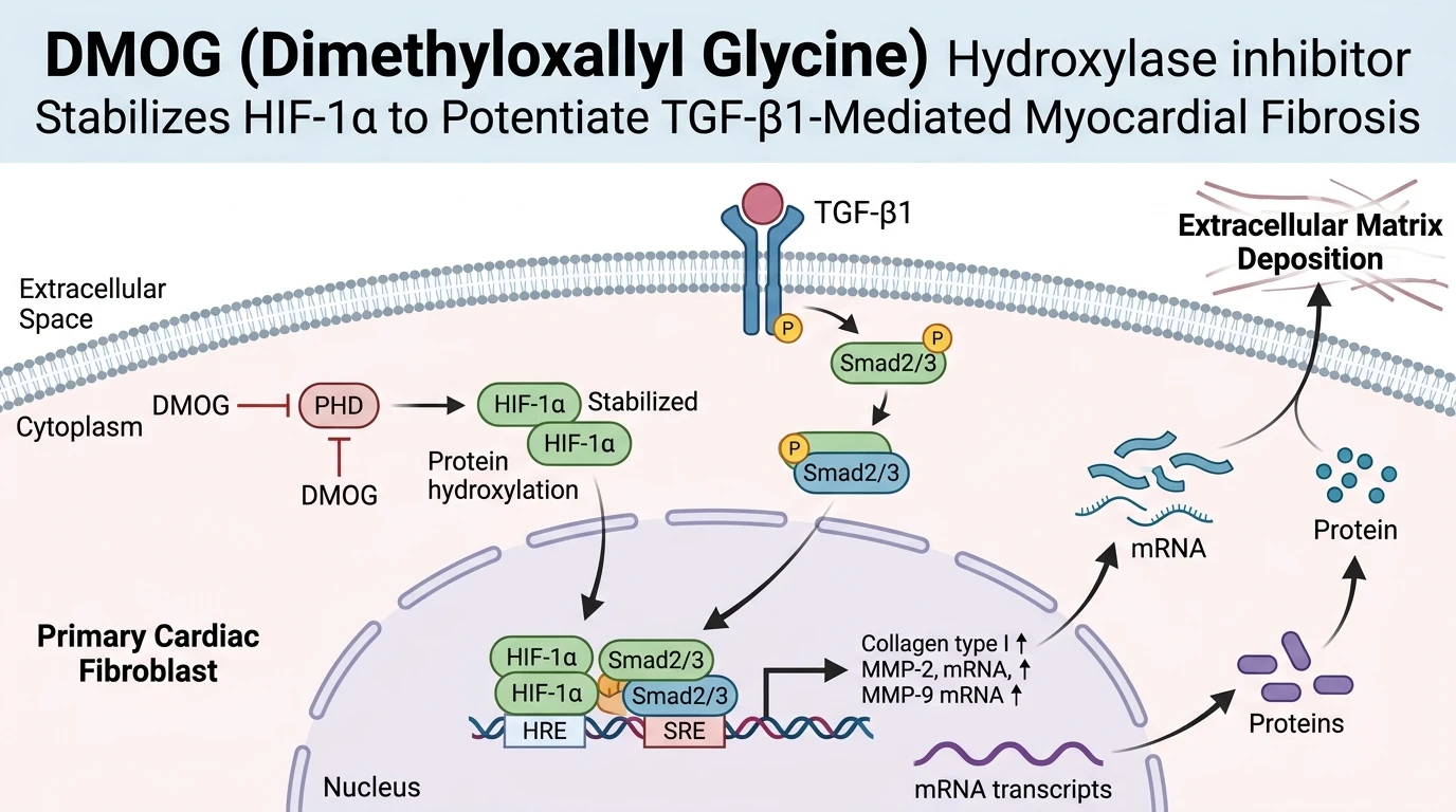
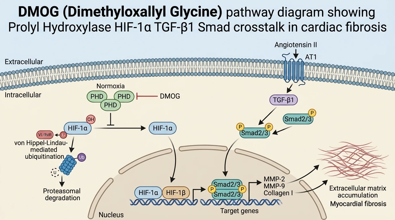
Hypoxia-inducible factor 1-alpha (HIF-1α) serves as a primary transcriptional regulator of cellular adaptation to reduced oxygen availability. Under normoxic conditions, specific prolyl hydroxylase domain (PHD) enzymes catalyze the hydroxylation of proline residues (Pro402 and Pro564) within the oxygen-dependent degradation domain of HIF-1α. This hydroxylation targets HIF-1α for recognition by the von Hippel-Lindau (pVHL) E3 ubiquitin ligase complex, leading to rapid polyubiquitination and 26S proteasomal degradation. Small molecule intervention using a competitive target like DMOG disrupts this homeostatic degradation mechanism by mimicking 2-oxoglutarate, the essential co-substrate for PHD catalytic activity.
Inhibiting PHD enzymes with DMOG stabilizes cellular HIF-1α levels, permitting its nuclear translocation and heterodimerization with HIF-1β. The resulting transcriptionally active HIF-1 complex binds to hypoxia response elements (HREs) across promoter regions of target genes involved in metabolic reprogramming, microvascular adaptation, and extracellular matrix (ECM) reorganization. In cardiac tissue contexts, Angiotensin II (Ang II) signaling through the Angiotensin II Type 1 (AT1) receptor acts in concert with hypoxic cues to stimulate endogenous HIF-1α synthesis and activate downstream transforming growth factor-beta 1 (TGF-β1) pathways.
Activation of the AT1 receptor initiates a signaling cascade that elevates intracellular reactive oxygen species and downstream kinase cascades, enhancing TGF-β1 expression. Secreted TGF-β1 binds to its heteromeric serine/threonine kinase receptor complex, inducing phosphorylation of receptor-regulated Smad proteins (Smad2 and Smad3). Phosphorylated Smad2/3 recruits Smad4 to form a transcriptional complex that migrates to the nucleus. When HIF-1α is simultaneously stabilized via PHD inhibition, nuclear HIF-1α physically and functionally interacts with the Smad protein complex. Structural domain analysis using protein domain family annotation tools reveals conserved interaction motifs within hydroxylase and PAS domains that facilitate co-regulator assembly. This synergistic convergence on gene promoters amplifies the transcription of key structural and remodeling proteins, including Collagen Type I, Matrix Metalloproteinase-2 (MMP-2), and Matrix Metalloproteinase-9 (MMP-9).
PHD enzymes belong to the iron- and 2-oxoglutarate-dependent dioxygenase superfamily. DMOG functions as a cell-permeable ester prodrug that hydrolyzes intracellularly into dimethyloxalylglycine, a structural analogue of 2-oxoglutarate. By competing with 2-oxoglutarate for the active site iron binding pocket of PHD1, PHD2, and PHD3, DMOG inhibits catalytic iron coordination and prevents oxygen-dependent proline hydroxylation.
In primary myocardial cell assays, exposure to hypoxia or Ang II treatment leads to sustained HIF-1α accumulation, accompanied by elevated TGF-β1 secretion. Elevated matrix metalloproteinase activity—specifically MMP-2 and MMP-9—facilitates extracellular matrix cleavage, creating space for structural collagen deposition while generating bioactive matrix fragments that further stimulate fibroblast differentiation into myofibroblasts. Myofibroblasts express elevated alpha-smooth muscle actin (α-SMA) and synthesize massive quantities of Collagen I and III fibers.
When AT1 receptors are pharmacologically blocked with valsartan, Ang II-driven TGF-β1 release and HIF-1α stabilization drop significantly. Consequently, cellular MMP-2 and MMP-9 expression levels return near baseline. However, introducing DMOG bypasses receptor-level inhibition. By selectively blocking PHD enzymatic turn-over, DMOG preserves HIF-1α protein integrity regardless of AT1 receptor state. Once stabilized, nuclear HIF-1α binds co-activators p300/CBP and cooperates with TGF-β1-induced Smad3 at adjacent DNA binding motifs, maximizing transcription of fibrotic effector genes. This molecular synergy explains why dual stimulation with DMOG and TGF-β1 produces a pronounced increase in collagen deposition in vitro and ex vivo.
Pharmacological Crosstalk and Fibrotic Effector Regulation
Copy code to add this image to your page
DMOG acts as a competitive prolyl hydroxylase inhibitor, preventing normoxic degradation of HIF-1α and driving its cytoplasmic accumulation. Stabilized HIF-1α translocates to the nucleus, where it acts synergistically with downstream effectors of AT1 receptor activation—specifically the TGF-β1/Smad2/3 pathway. This dual pathway engagement markedly amplifies transcriptional induction of matrix metalloproteinases (MMP-2 and MMP-9) and Collagen I. Consequently, DMOG-mediated HIF-1α stabilization directly accelerates extracellular matrix deposition and fibrotic remodeling in cardiac tissues following ischemic or hypertensive stress.
AT1 Receptor Blockade Suppresses HIF-1α and ECM Production
Angiotensin II stimulation elevates HIF-1α accumulation in primary cardiac fibroblasts. Pharmacological receptor blockade using valsartan significantly reduced downstream AT1 receptor signaling pathways, causing a measurable decrease in cellular TGF-β1 synthesis. Total collagen synthesis dropped sharply within twenty-four hours. These empirical data confirm that AT1 receptor activation contributes directly to normoxic HIF-1α accumulation, whereas receptor antagonists effectively attenuate fibrotic marker expression in vitro.
Targeting Hydroxylases Restores Fibrotic Effector Gene Expression
Prolyl hydroxylase activity ceased following compound administration. Treatment with DMOG (Dimethyloxallyl Glycine) Hydroxylase inhibitor bypassed AT1 receptor blockade, driving rapid HIF-1α accumulation within both cytoplasmic and nuclear compartments. Western blot analysis confirmed nuclear translocation of stabilized HIF-1α. Restored HIF-1α expression partially recovered MMP-2 and MMP-9 protein levels, which subsequently enhanced Collagen I secretion in primary cardiac myofibroblast cultures under tested assay conditions.
Synergistic HIF-1α and TGF-β1 Crosstalk Accelerates Collagen Synthesis
Dual pathway activation accelerated matrix protein deposition. Co-administration of DMOG and exogenous TGF-β1 produced elevated collagen accumulation that significantly exceeded individual responses observed in primary cardiac cell models. Smad2 and Smad3 phosphorylation remained sustained throughout culture. Physical interaction between nuclear HIF-1α and active Smad complexes amplified transcriptional induction, proving that prolyl hydroxylase inhibition acts synergistically with TGF-β signaling to drive myocardial remodeling.