TEPP-46 (ML265) Allosterically Activates Pyruvate Kinase to Modulate Pancreatic Alpha Cell Calcium Dynamics
Copy code to add this image to your page
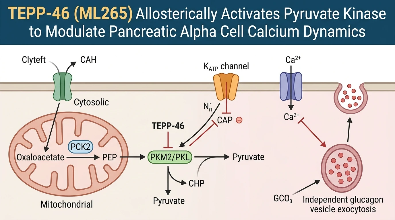
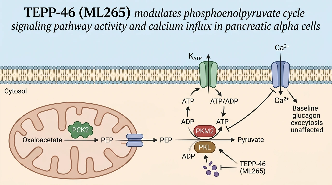
Pancreatic α-cells regulate glucose counter-regulation through the synthesis and secretion of glucagon. Intracellular nutrient sensing in these cells relies on tightly regulated metabolic cycles that couple mitochondrial activity to ion channel gating at the plasma membrane. Central to this metabolic architecture is the mitochondrial phosphoenolpyruvate (PEP) cycle. Mitochondria generate oxaloacetate, which is converted into PEP by mitochondrial phosphoenolpyruvate carboxykinase (PCK2). Cytosolic export of PEP provides a localized pool of substrate for pyruvate kinase isoforms, primarily PKM2 and PKL. Pyruvate kinase catalyzes the conversion of PEP and ADP into pyruvate and ATP. This localized ATP production elevates the sub-plasma membrane ATP/ADP ratio, triggering the closure of ATP-sensitive potassium (KATP) channels, membrane depolarization, and subsequent influx of extracellular Ca2+ through voltage-gated calcium channels.
Targeting this pathway, TEPP-46 (ML265) serves as a selective, small-molecule allosteric activator of pyruvate kinase. Structurally, TEPP-46 binds at the subunit interaction interface of PKM2 and PKL tetramers. This binding stabilizes the active tetrameric conformation and prevents monomerization or dimerization, thereby driving the enzyme toward a high-affinity state for PEP. In primary pancreatic α-cell preparations, application of TEPP-46 at 10 µM dramatically alters nutrient-induced intracellular signaling. When α-cells are challenged with amino acid mixtures, allosteric activation by TEPP-46 causes a rapid depletion of cytosolic PEP pools and accelerates ATP turnover. Surprisingly, this pharmacologically driven acceleration of pyruvate kinase activity results in the near-total abolition of amino acid-stimulated Ca2+ influx.
To establish whether this Ca2+ suppression stems specifically from PEP cycle kinetics, complementary genetic knockout models were analyzed. Mice harboring alpha-cell-specific deletions of pyruvate kinase (PKM1/2-αKO) or mitochondrial PEP carboxykinase (PCK2-αKO) display opposite electrophysiological phenotypes. Loss of PCK2 or PKM isoforms blunts local PEP production or consumption, leading to enhanced intracellular Ca2+ responses following amino acid exposure. Patch-clamp electrophysiology demonstrates altered baseline membrane potential and sustained KATP channel activity in these knockouts. These observations indicate that an operational mitochondrial PEP cycle acts as an intrinsic metabolic limiter, buffering the magnitude of Ca2+ signals during nutrient abundance.
The metabolic compartmentalization of pyruvate kinase isoforms within pancreatic islet cells contributes to distinct functional behaviors between α-cells and β-cells. While β-cells rely heavily on PKM2 activation to drive insulin secretion via KATP channel closure, α-cells express a co-existing population of PKM2 and PKL isoforms. The presence of PKL provides a distinct kinetic threshold, allowing α-cells to tune their metabolic response across varying concentrations of amino acids and glucose. When TEPP-46 binds to PKM2 and PKL, it locks both isoforms in their active conformations, overriding native regulatory mechanisms such as allosteric inhibition by phenylalanine or regulatory phosphorylation. This constant hyper-activation accelerates PEP turnover, preventing the transient accumulation of high-energy phosphate intermediates near KATP channels needed to sustain prolonged Ca2+ spikes.
Crucially, these distinct Ca2+ dynamics do not translate into altered hormone output. Pharmacological activation with TEPP-46 does not impair or enhance amino acid-induced glucagon secretion. Similarly, genetic deletion of PCK2 or PKM1/2 leaves baseline and stimulated glucagon release fully intact. In classic endocrine paradigms, intracellular Ca2+ elevation is considered an absolute prerequisite for dense-core vesicle exocytosis. However, in pancreatic α-cells under these tested assay conditions, amino acid-stimulated glucagon exocytosis proceeds independently of PEP cycle-mediated Ca2+ entry. TEPP-46 (ML265) thus serves as a valuable chemical probe, uncoupling sub-membrane Ca2+ flux from exocytotic machinery in α-cell stimulus-secretion coupling.
Pyruvate Kinase Activation and Alpha-Cell Bioenergetics
Copy code to add this image to your page
TEPP-46 (ML265) acts as an allosteric activator of pyruvate kinase isoforms PKM2 and PKL. By stabilizing the active tetrameric enzyme state, TEPP-46 accelerates cytosolic phosphoenolpyruvate (PEP) clearance. In pancreatic α-cells, this pharmacological activation severely attenuates amino acid-stimulated Ca2+ influx through altered KATP channel regulation. However, amino acid-induced glucagon secretion remains unaffected, demonstrating a mechanistic divergence between PEP cycle-mediated Ca2+ dynamics and exocytosis.
Allosteric Activation Suppresses Nutrient-Induced Calcium Influx
Pyruvate kinase converts cytosolic PEP into pyruvate while generating ATP near the plasma membrane. Administration of TEPP-46 at 10 µM selectively stabilizes the active tetrameric conformation of PKM2 and PKL. This pharmacological engagement markedly alters intracellular signaling events in pancreatic α-cells during amino acid stimulation. Under baseline conditions, amino acids induce membrane depolarization and robust Ca2+ entry. In the presence of the activator, this Ca2+ influx is nearly abolished. Short-term kinetic measurements confirm rapid PEP depletion. Consequently, local ATP/ADP ratios at the inner leaf of the plasma membrane fail to sustain KATP channel closure. Genetic knockouts of PKM1/2 or PCK2 produce opposing electrophysiological phenotypes, elevating cytosolic Ca2+ spikes. Thus, allosteric activation of pyruvate kinase acts as an effective brake on nutrient-induced electrical activity within this specific cellular model.
Glucagon Exocytosis Functions Independently of PEP Cycle Modulation
Endocrine dogma tightly couples intracellular Ca2+ elevation to peptide hormone release. However, experiments in isolated pancreatic α-cells demonstrate a clear divergence between Ca2+ dynamics and hormone output. Neither genetic disruption of PCK2 nor PKM1/2 deletion alters amino acid-stimulated glucagon secretion. Similarly, treatment with TEPP-46 confirmed this decoupling in primary islets. Glucagon output remained fully intact despite the profound reduction in Ca2+ influx. These observations indicate that amino acid sensing in α-cells recruits redundant pathways to drive vesicle exocytosis—possibly involving direct amino acid transport or cAMP-mediated priming. Observations in these in vitro models highlight a distinct separation between metabolic ion channel regulation and secretory machinery.