XMU-MP-1 MST Kinase Inhibition Drives G2/M Arrest in Hematologic Malignancies
Copy code to add this image to your page
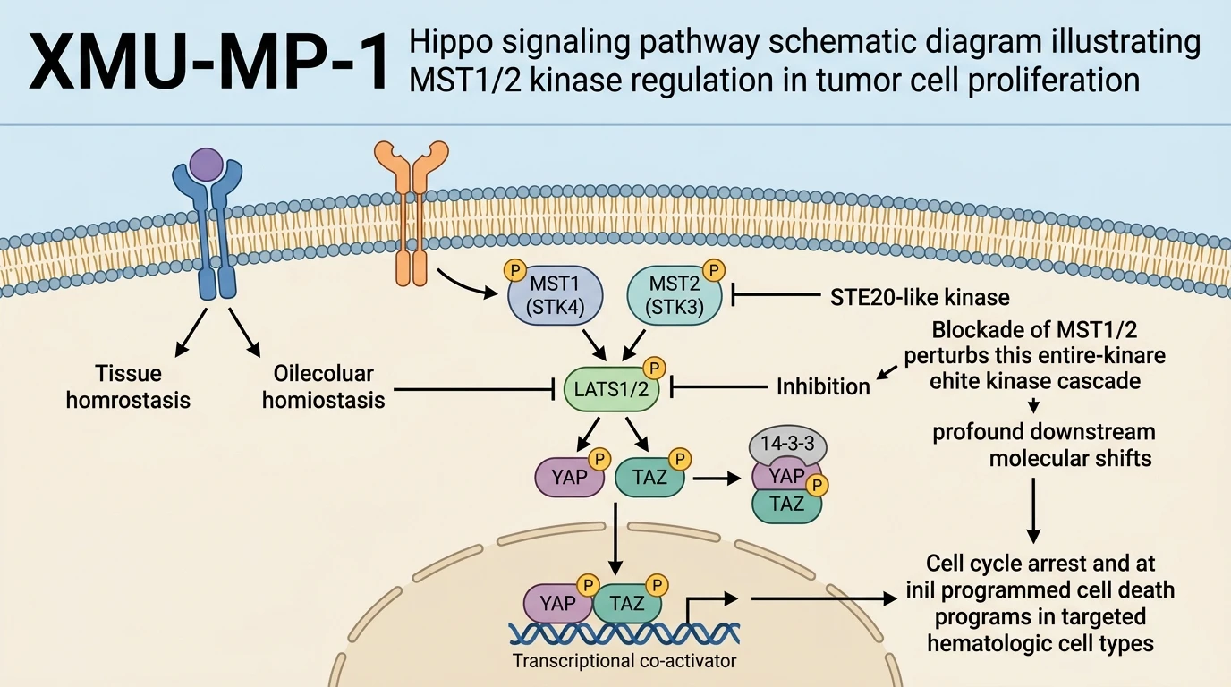
Targeting central enzymatic nodes within developmental signaling pathways offers a specialized vector for oncology therapeutics. The Hippo pathway core cassette, anchored by the mammalian sterile 20-like kinases MST1 (STK4) and MST2 (STK3), regulates cellular proliferation, organ size, and apoptosis. Pharmacological evaluation of small-molecule MST1/2 kinase inhibitors demonstrates striking context-dependent cytotoxic profiles across distinct neoplastic lineages. Among these targeted agents, XMU-MP-1 operates as a selective ATP-competitive inhibitor of MST1/2 kinases, disrupting homeostatic Hippo kinase cascades and altering downstream signal transduction.
Empirical evaluation across diverse human cancer cell lines highlights a pronounced lineage specificity. In vitro viability assays show that administration of XMU-MP-1 suppresses cell proliferation across B- and T-cell hematologic tumor models, including Namalwa, Raji, Ramos, Jurkat, and Daudi cell lines. Conversely, tested solid tumor models—such as breast cancer cell lines—maintain viability under equivalent dosing regimens. This observed differential sensitivity suggests that specific hematologic malignancies rely heavily on intact MST1/2 signaling networks for survival. To explore targeted molecular interactions, researchers routinely reference kinase domain sequence annotations to contextualize ATP-binding pocket mechanics and structural selectivity across kinase subfamilies.
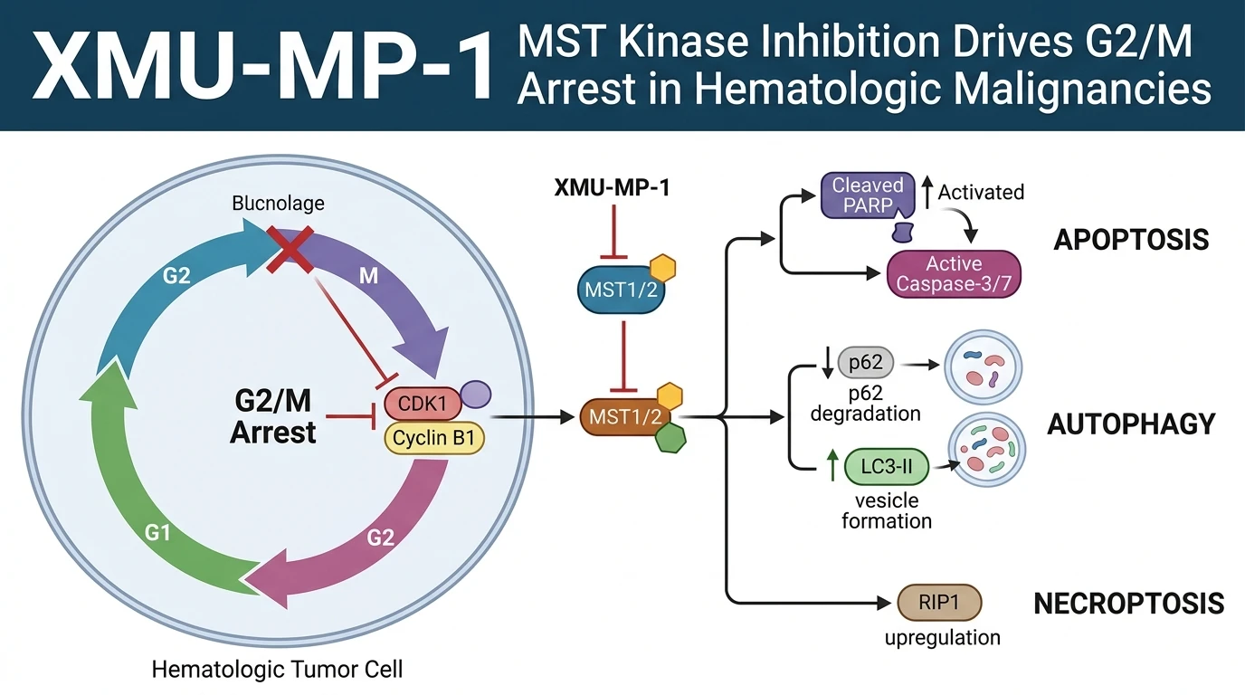
Mechanistic dissection of cell cycle dynamics reveals that target engagement yields substantial phenotypic changes prior to cell death. Flow cytometry profiling indicates that inhibitor treatment induces G2/M phase cell cycle arrest in sensitive hematologic lines. Biochemical analysis demonstrates a corresponding down-regulation of key cell cycle regulators, including E2F transcription factors, cell division cycle (CDC) proteins, cyclins, and cyclin-dependent kinases (CDKs). By halting the G2/M transition, the inhibitor prevents clonal expansion and destabilizes ongoing mitotic fidelity.
Beyond mitotic arrest, sustained kinase suppression triggers multiple redundant death programs. Rather than relying on a single cytotoxic modality, treated cells display biochemical hallmarks of apoptosis, autophagy, and necroptosis. Apoptotic engagement is evidenced by marked elevation of caspase 3/7 enzymatic activity and downstream poly(ADP-ribose) polymerase (PARP) cleavage. Concurrently, autophagic flux increases, as marked by p62 protein degradation and concomitant LC3-II accumulation. Analysis of necroptotic nodes reveals significant upregulation of receptor-interacting protein kinase 1 (RIP1). This multi-modal activation of cell death pathways indicates that targeting MST1/2 disrupts cellular baseline homeostasis across parallel survival axes.
Pharmacological Characterization and Multi-Modal Cytotoxic Dynamics
Copy code to add this image to your page
Selective pharmacological inhibition of MST1/2 kinases by XMU-MP-1 alters core Hippo signaling cascades, inducing marked cytotoxicity in B- and T-cell hematologic tumor models while sparing non-hematologic tissues. Target engagement drives G2/M phase cell cycle arrest through the systematic down-regulation of E2Fs, CDCs, cyclins, and CDKs. Furthermore, sustained MST1/2 blockade triggers active cell death across three distinct pathways: apoptosis (caspase 3/7 activation, PARP cleavage), autophagy (p62 degradation, LC3-II accumulation), and necroptosis (RIP1 upregulation).
Hematologic Lineage Sensitivity Under Selective Kinase Blockade
Target validation studies confirm marked lineage-specific responses following MST suppression. Application of XMU-MP-1 MST inhibitor compounds selectively reduces viable cell counts in panel assays of B- and T-cell lines. Namalwa, Raji, Ramos, Jurkat, and Daudi lines displayed pronounced growth inhibition at nanomolar to low-micromolar concentrations. In contrast, parallel evaluations in breast adenocarcinoma models demonstrated negligible cytotoxicity. Cellular reliance on baseline Hippo pathway activity appears markedly higher in hematologic lineages. Proliferation assays confirmed rapid growth arrest within 24 hours of exposure. Selective toxicity remained stable across varying serum concentrations in cultured media.
G2/M Phase Arrest Driven by Mitotic Regulator Suppression
Cellular cycle progression requires tightly synchronized expression of cyclin-CDK complexes. Flow cytometric analysis reveals that drug exposure locks treated hematologic cells in the G2/M boundary. Downstream transcriptomic and western blot analyses document coordinated down-regulation of E2F transcription factors and CDC family phosphatases. Mitotic cyclins and their partner CDKs showed reduced expression levels post-treatment. This concerted depletion of cell cycle machinery prevents spindle assembly completion. Mitotic arrest persisted prior to the emergence of irreversible cytotoxic markers. The arrest phase provides a critical window where damaged cells transition toward active programmed cell death pathways.
Simultaneous Engagement of Apoptotic Autophagic and Necroptotic Nodes
Prolonged G2/M arrest initiates converging cell death cascades. Treatment with XMU-MP-1 MST inhibitors rapidly activates executor caspases 3 and 7. Cleavage of PARP confirmed functional apoptotic execution in a dose-dependent manner. Simultaneously, autophagic processing intensified across treated cohorts. Protein levels of p62 decreased significantly while LC3-I converted rapidly to lipidated LC3-II. Necroptotic machinery also responded directly to targeted inhibition. RIP1 protein levels rose, indicating engagement of programmed necrosis alongside classical apoptosis. This concurrent execution across three distinct death programs reduces the probability of acquired single-pathway resistance.