MCC950 Inhibits NLRP3 Inflammasome via NACHT Domain Binding in Diabetic Retinopathy
Copy code to add this image to your page
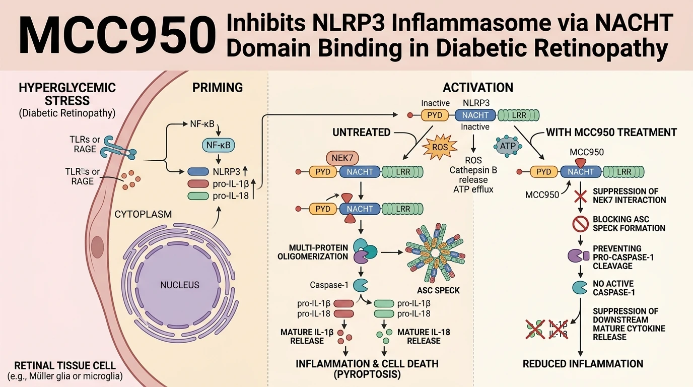
Intracellular pattern recognition receptors act as sensory hubs for metabolic derangements. Among these sensor proteins, the NACHT, LRR, and PYD domains-containing protein 3 (NLRP3) inflammasome functions as a primary driver of sterile vascular inflammation in diabetic retinopathy (DR). In retinal microvascular endothelial cells and retinal pigment epithelial cells exposed to sustained elevated glucose levels, intracellular reactive oxygen species (ROS) accumulate rapidly. High ROS levels trigger up-regulation of NIMA-related kinase 7 (NEK7), an essential serine/threonine kinase that serves as a licensing cofactor for NLRP3 activation. NEK7 binds directly to the central NACHT domain of NLRP3, inducing a conformational rearrangement necessary for ASC (apoptosis-associated speck-like protein containing a CARD) recruitment. Subsequent ASC oligomerization recruits pro-Caspase-1, driving auto-proteolytic cleavage into active Caspase-1 (Cleaved Caspase-1). Active Caspase-1 processes pro-interleukin-1β (pro-IL-1β) and pro-interleukin-18 (pro-IL-18) into their bioactive mature forms, which perpetuate microvascular breakdown, blood-retinal barrier leakage, and apoptotic death of neuroretinal cells.
Targeting this multi-protein complex requires specific structural intervention. MCC950 is a potent sulfonylurea-based small molecule designed to directly inhibit canonical and non-canonical NLRP3 activation. Biochemical assays confirm that inhibition of NLRP3 inflammasome signaling by MCC950 occurs through high-affinity direct binding to the ATP-hydrolysis NACHT domain of NLRP3. Direct binding traps NLRP3 in a closed, inactive conformation, stericly hindering the association between NEK7 and the NLRP3 NACHT domain. Without NEK7-NACHT domain stabilization, supramolecular assembly of the ASC speck complex is effectively blocked.
In preclinical rodent models of streptozotocin (STZ)-induced type 2 diabetes mellitus (T2DM), sustained hyperglycemia induces substantial oxidative stress and histopathological damage in retinal tissue layers. Intravitreal administration of MCC950, particularly at an optimized concentration of 1 mM, exerts pronounced local anti-inflammatory and retinoprotective effects. Intravitreal delivery bypasses systemic exposure limitations while delivering effective local drug concentrations directly to the posterior segment of the eye. Experimental evaluation reveals that MCC950 administration suppresses ROS generation within diabetic retinal tissue. Reduced ROS levels downregulate cellular expression of NEK7, interrupting a pathogenic feed-forward loop where ROS amplifies NEK7 expression and subsequent inflammasome priming.
At the protein level, 1 mM MCC950 therapy significantly downregulates total expression levels of NLRP3, NEK7, and ASC within the diabetic retina. This transcriptional and post-translational suppression directly diminishes Cleaved Caspase-1 abundance. Consequently, retinal tissue concentrations of mature pro-inflammatory cytokines IL-1β and IL-18 are markedly reduced. Retinal histopathology demonstrates structural preservation of the inner nuclear layer and ganglion cell layer, accompanied by a quantifiable reduction in terminal deoxynucleotidyl transferase dUTP nick end labeling (TUNEL)-positive apoptotic neuroretinal cells.
Pharmacodynamic characterization in STZ-induced rat models demonstrates dose-dependent suppression of inflammatory markers following intravitreal injection. While lower micromolar concentrations demonstrate partial attenuation of Caspase-1 activation, a 1 mM intravitreal dose achieves near-complete blockade of retinal IL-1β maturation without triggering ocular toxicity or intraocular pressure elevation. Western blot analyses of retinal tissue lysates show a marked decline in the ratio of Cleaved Caspase-1 to pro-Caspase-1 following MCC950 intervention. Immunohistochemical staining further confirms reduced ASC speck formation within the retinal ganglion cell layer and inner plexiform layer. Together, these biochemical markers correlate directly with reduced microvascular permeability, decreased acellular capillary counts, and functional preservation of retinal electroretinogram responses under scotopic and photopic testing conditions.
The therapeutic selectivity of MCC950 is distinct among anti-inflammatory agents. Unlike broad-spectrum anti-inflammatory agents or pan-caspase inhibitors, MCC950 selectively targets NLRP3 without disrupting AIM2, NLRC4, or NLRP1 inflammasome pathways. This target selectivity maintains fundamental host innate immune defenses against pathogen challenge while neutralizing chronic, sterile metabolic inflammation within hyper-glycemic ocular tissue. These empirical observations establish the ROS-NEK7-NLRP3 signaling axis as an actionable target for mitigating microvascular and neurodegenerative pathologies characteristic of diabetic retinopathy.
Pharmacological Evaluation of MCC950 in Retinal Tissue
Copy code to add this image to your page
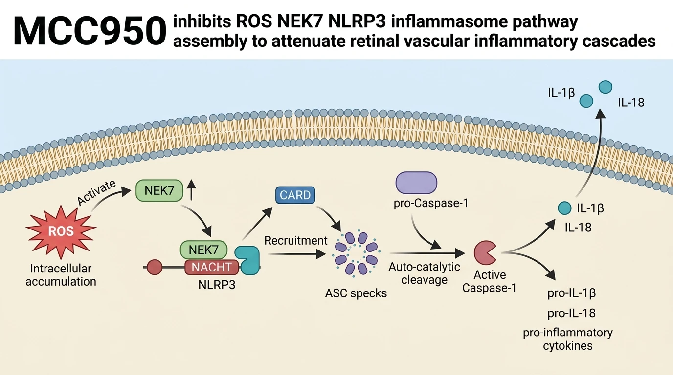
MCC950 is a selective, small-molecule inhibitor of the NLRP3 inflammasome that directly binds to the central NACHT domain of NLRP3. By physically blocking the NACHT domain, MCC950 prevents the essential interaction between NEK7 and NLRP3 required for inflammasome oligomerization. This structural blockade inhibits ASC speck formation and abolishes pro-Caspase-1 autocatalytic cleavage. Downstream processing of mature IL-1β and IL-18 cytokines is significantly diminished. Concurrently, MCC950 suppresses ROS-mediated NEK7 expression, interrupting positive feedback amplification loops in retinal tissue under chronic hyper-glycemic stress.
Direct NACHT Domain Binding Blocks NEK7 Assembly
MCC950 binds directly to the nucleotide-binding domain of NLRP3. This interaction prevents ATP hydrolysis within the NACHT domain; consequently, the receptor remains locked in an inactive state. Physical occlusion of the binding interface disrupts the assembly of the NEK7-NLRP3 complex. In the absence of stable NEK7 association, NLRP3 cannot undergo the self-oligomerization required for ASC recruitment. ASC speck formation ceases completely. Downstream cleavage of pro-Caspase-1 is thus blocked at the initial biochemical step. In STZ-induced T2DM rat retinas, 1 mM intravitreal MCC950 restored basal cytokine profiles. Cleaved Caspase-1 levels dropped significantly following treatment. Mature IL-1β and IL-18 concentrations returned toward non-diabetic baselines within retinal tissue homogenates.
Intravitreal Delivery Mitigates Hyperglycemia-Induced Retinal Apoptosis
Hyperglycemia generates excess intracellular ROS within neuroretinal cells. High ROS concentrations upregulate NEK7 protein expression and promote oxidative cell death. Intravitreal administration of MCC950 attenuates local oxidative stress markers. ROS levels declined rapidly in treated retinal tissue. Decreased ROS concentrations correlated with marked downregulation of NEK7 and ASC expression. Histopathological analysis confirmed structural preservation of the outer nuclear and inner plexiform layers. TUNEL assays revealed a sharp decline in apoptotic neuroretinal cells after treatment. Neuroretinal structural integrity was maintained under chronic hyper-glycemic stress. These experimental observations remain bounded by the specific STZ-induced rodent model evaluated.