Ac-CoA Synthase Inhibitor1 (ACSS2 inhibitor) dampens mTORC1 signaling to attenuate diabetic nephropathy
Copy code to add this image to your page
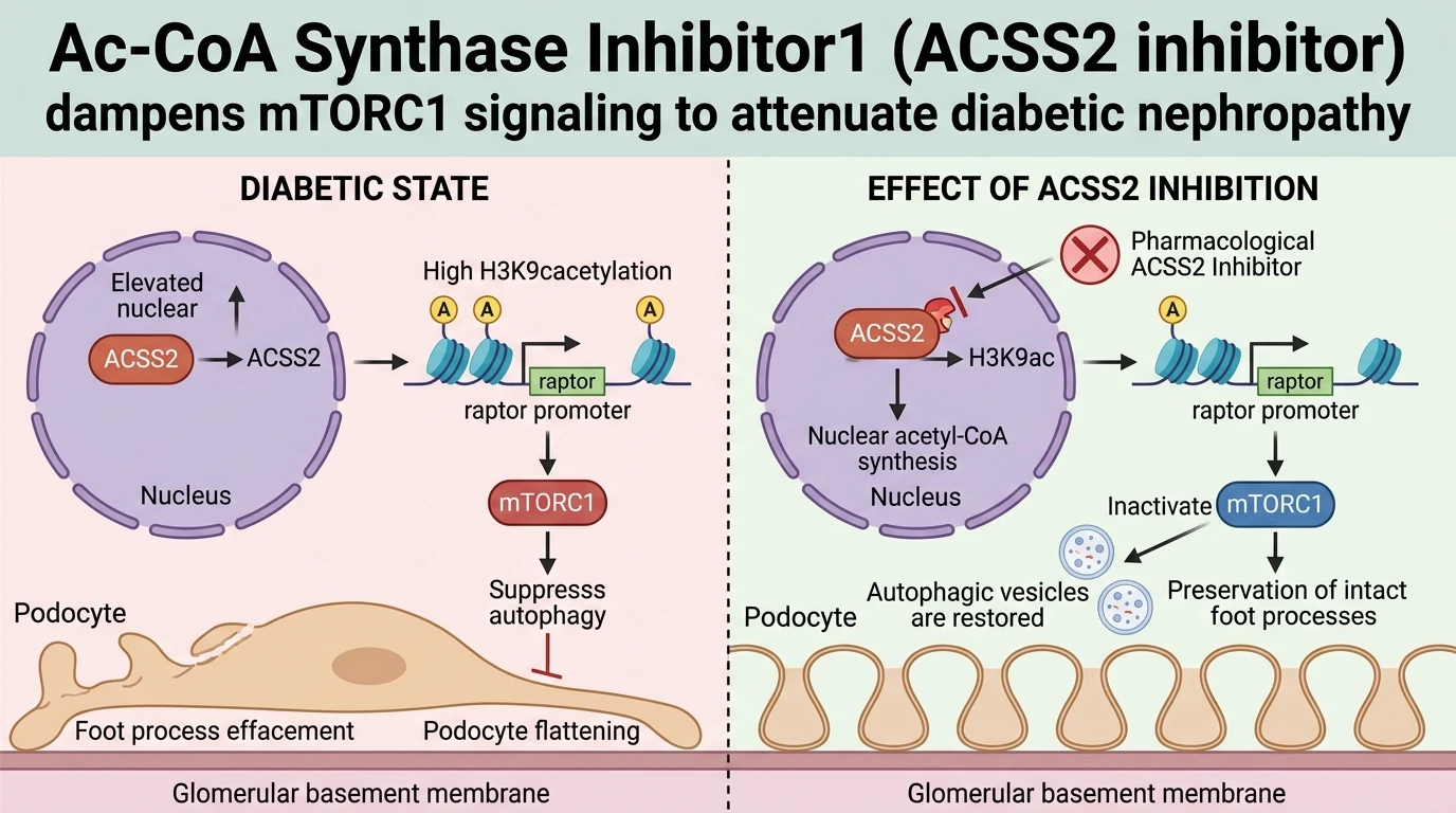
In diabetic nephropathy, sustained hyperglycemia alters metabolic enzyme expression within glomerular podocytes. Acetyl-CoA synthetase 2 (ACSS2) functions as a primary nucleocytosolic enzyme converting free acetate into acetyl-CoA. Quantitative transcriptomic and immunohistochemical profiling reveals marked ACSS2 upregulation in renal biopsies from diabetic nephropathy patients as well as streptozotocin-induced diabetic mice. This upregulation directly correlates with serum creatinine accumulation and urinary albumin excretion. Elevated ACSS2 expression increases the local nuclear supply of acetyl-CoA. Consequently, histone acetyltransferases utilize this metabolic metabolite pool to alter chromatin accessibility across specific gene loci.
Epigenetic profiling demonstrates that elevated ACSS2 preferentially enriches histone H3 lysine 9 acetylation (H3K9ac) at the promoter region of regulatory-associated protein of mTOR (raptor). Raptor serves as an essential structural subunit of mammalian target of rapamycin complex 1 (mTORC1). Hyperacetylation at the raptor promoter facilitates transcriptional machinery recruitment, driving elevated raptor protein expression. Heightened raptor assembly activates downstream mTORC1 kinase activity in podocytes exposed to high-glucose conditions. Activated mTORC1 phosphorylates Unc-51 like autophagy activating kinase 1 (ULK1) at inhibitory residues, thereby suppressing autophagosome formation. Podocytes rely heavily on basal autophagy to maintain structural integrity and filtration barrier function. Autophagic suppression causes accumulation of damaged mitochondria, increases oxidative stress, and triggers podocyte phenotypic transition toward a mesenchymal state characterized by loss of synaptopodin and gain of desmin expression.
Application of pharmacological targeting of epigenetic regulators using Ac-CoA Synthase Inhibitor1 (ACSS2 inhibitor) disrupts this pathogenic cascade. Small-molecule inhibition of ACSS2 selectively depletes nuclear acetyl-CoA pools in diabetic podocytes without altering baseline cytoplasmic acetyl-CoA levels required for fatty acid synthesis. Reduced nuclear acetyl-CoA levels lead to rapid deacetylase-mediated clearance of H3K9ac at the raptor promoter. Downregulation of raptor expression dismantles active mTORC1 complexes, releasing ULK1 from inhibitory phosphorylation. Restored autophagic flux restores cellular proteostasis, mitigates cellular swelling, and prevents foot process effacement under high-glucose challenge in vitro and in vivo.
Epigenetic mTORC1 Modulation and Autophagic Restoration in Podocytes
Copy code to add this image to your page
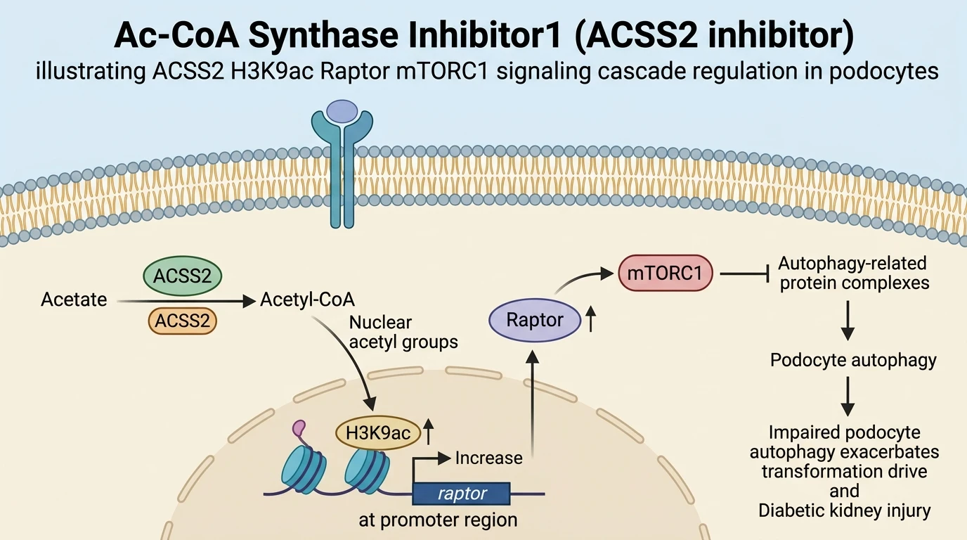
Ac-CoA Synthase Inhibitor1 (ACSS2 inhibitor) selectively blocks nuclear acetyl-CoA synthesis mediated by ACSS2 in diabetic podocytes. Decreased chromatin-localized acetyl-CoA lowers H3K9 acetylation at the promoter of raptor, downregulating raptor expression. Reduced raptor levels suppress hyperactive mTORC1 signaling, thereby disinhibiting ULK1-driven autophagy pathways. Rescued autophagic clearance limits podocyte phenotypic transformation, reduces inflammatory signaling, and halts progressive glomerular injury in diabetic nephropathy.
ACSS2 Epigenetically Activates Raptor via Promoter H3K9 Acetylation
High glucose drives nuclear accumulation of ACSS2 in cultured podocytes. Local conversion of acetate generates localized acetyl-CoA microdomains near chromatin. Chromatin immunoprecipitation assays confirmed marked enrichment of H3K9ac at the raptor promoter region under hyper-glycemic stress. Increased histone acetylation destabilizes nucleosomes and promotes RNA polymerase II recruitment. Consequently, raptor mRNA and protein levels rose significantly. Elevated raptor expression accelerated assembly of functional mTORC1 complexes. This nuclear metabolic signaling axis directly bridges cytosolic nutrient excess with epigenetic transcription.
Targeting ACSS2 Suppresses mTORC1 to Restore Podocyte Autophagy
Pharmacological inhibition using an Ac-CoA inhibitor attenuates raptor transcript abundance in treated podocytes. Depletion of nuclear acetyl-CoA rapidly diminished promoter-associated H3K9ac marks within twelve hours. Reduced raptor levels blunted mTORC1 activity, lowering p-S6K1 and p-ULK1 phosphorylation. Autophagic initiation resumed immediately. LC3-II convertibility increased substantially, accompanied by accelerated p62 clearance. Autophagosome formation was verified via transmission electron microscopy. Restored autophagic clearance reduced cytoplasmic protein aggregation and mitigated podocyte apoptosis under ambient high-glucose conditions.
In Vivo Amelioration of Albuminuria and Glomerular Histopathology
Administration of the small-molecule Ac-CoA inhibitor improved renal outcomes in diabetic mouse models. Urinary albumin-to-creatinine ratios dropped significantly after four weeks of daily therapy. Histopathological analysis confirmed reduction in mesangial matrix expansion and basement membrane thickening. Podocyte density within glomeruli remained preserved relative to vehicle-treated diabetic controls. Synaptopodin expression recovered, confirming preservation of foot process architecture. These physiological changes demonstrate that targeted metabolic-epigenetic modulation protects renal filtration integrity during diabetic stress.