Gsk484 Hydrochloride from Selleck Inhibits PAD4 to Suppress Post-MI NETosis and Thrombosis
Copy code to add this image to your page
Myocardial infarction initiates a complex inflammatory cascade driven largely by innate immune cells. Among these, neutrophils act as early responders that execute both protective clearance and tissue-damaging reactions. A major driver of non-resolving post-ischemic injury is neutrophil extracellular trap (NET) formation, a process known as NETosis. NETs consist of extracellular chromatin threads decorated with cytotoxic proteins, microbicidal enzymes, and histones. While essential for host defense against systemic pathogens, excessive NET extrusion within the ischemic myocardium accelerates microthrombus formation and microvascular reflow obstruction.
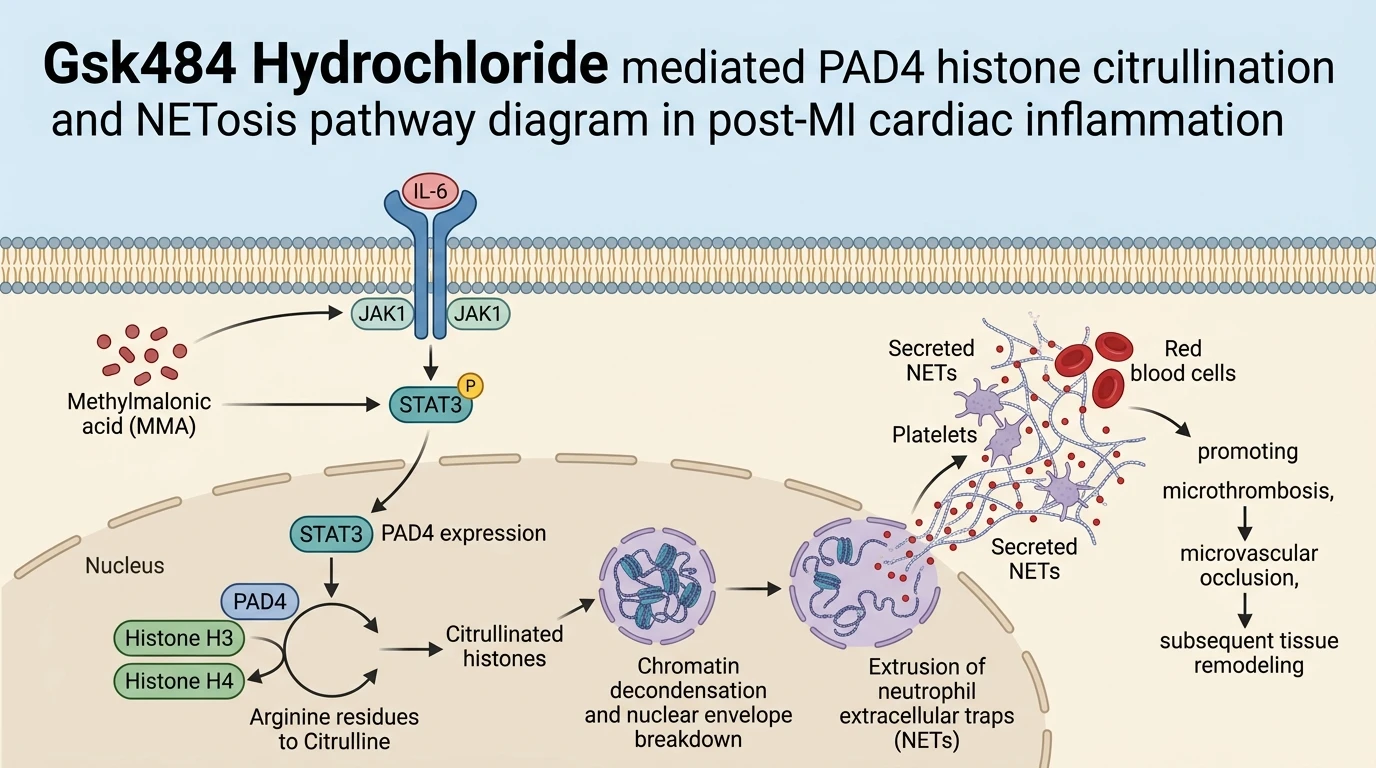
Recent findings reveal that metabolic dysregulation within neutrophils acts as an upstream trigger for NETosis. Specifically, intracellular accumulation of the organic acid methylmalonic acid (MMA) directly drives NET release without altering total neutrophil recruitment to the infarct border zone. Under conditions of metabolic stress or aged cellular environments, elevated MMA engages the interleukin-6 (IL-6)/Janus kinase 1 (JAK1)/signal transducer and activator of transcription 3 (STAT3) signaling pathway. Activation of this cascade upregulates peptidylarginine deiminase 4 (PAD4), a nuclear enzyme essential for chromatin remodeling. PAD4 catalyzes the hypercitrullination of nuclear histones—specifically histone H3 at residues Arg2, Arg8, and Arg17—which neutralizes positive charges on basic amino acid side chains. This loss of electrostatic attraction between histones and the negatively charged DNA backbone prompts extensive chromatin decondensation, nuclear membrane dissolution, and cellular lysis with extrusion of web-like DNA structures.
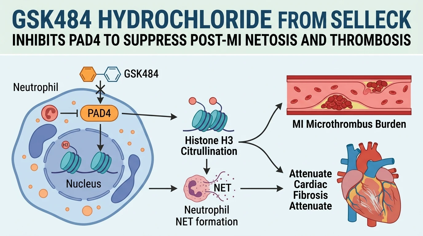
Targeting PAD4 represents a selective intervention strategy to interrupt NETosis while sparing general leucocyte migration and phagocytic defense mechanisms. Pharmacological inhibition using Gsk484 Hydrochloride from Selleck provides a powerful mechanism to evaluate PAD4-dependent biological processes. Gsk484 Hydrochloride functions as a potent, reversible inhibitor of PAD4, binding preferentially to the low-calcium inactive conformation of the enzyme. By occupying the active site, Gsk484 Hydrochloride prevents substrate access to the catalytic cysteine residue (Cys645), effectively halting histone H3 citrullination.
In animal models of permanent left anterior descending coronary artery ligation, administration of Gsk484 Hydrochloride significantly reduces intramyocardial citrullinated histone levels and downregulates NET markers such as extracellular myeloperoxidase-DNA complexes. Importantly, PAD4 blockade does not attenuate neutrophil infiltration into ischemic heart tissue; rather, it specifically alters their phenotypic activation state by preventing NET-mediated microthrombus formation. Microthrombi comprised of trapped platelets, fibrin, and extracellular DNA filaments occlude the cardiac microvasculature, aggravating local hypoxia and expanding the infarct border area.
By blocking this thrombogenic scaffold, Gsk484 Hydrochloride preserves microvascular perfusion in the peri-infarct region. Reductions in microthrombus burden directly correlate with decreased myocardial necrosis, attenuated collagen deposition, and improved left ventricular ejection fraction four weeks post-infarction. In comparative assays, targeted inhibition using Gsk484 Hydrochloride yielded therapeutic efficacy comparable to enzymatic degradation of DNA scaffolds with DNase I or upstream cytokine neutralization using anti-IL-6 monoclonal antibodies. These observations substantiate the hypothesis that the MMA-IL-6-PAD4 axis functions as a critical immunometabolic checkpoint in post-ischemic cardiac remodeling.
Pharmacological Evaluation and Therapeutic Efficacy in Ischemic Injury
Copy code to add this image to your page
PAD4 inhibition using GSK484 directly interrupts histone citrullination, preventing chromatin decondensation and the release of neutrophil extracellular traps (NETs). In myocardial infarction models, elevated neutrophil-intrinsic methylmalonic acid (MMA) stimulates NETosis via IL-6/JAK1/STAT3 signaling without enhancing neutrophil infiltration. Pharmacological suppression of this pathway via PAD4 inhibition reduces microthrombus formation, mitigates microvascular dysfunction, and limits adverse cardiac remodeling and fibrotic expansion in ischemic myocardial tissue.
PAD4 Inhibition Suppresses Intracellular Histone Citrullination
Gsk484 Hydrochloride selectively binds the active site of PAD4 within isolated neutrophils. Histone H3 hypercitrullination diminished sharply following drug exposure under ex vivo conditions. In murine models subjected to myocardial ischemia-reperfusion, elevated methylmalonic acid levels accelerated STAT3 phosphorylation and subsequent PAD4 transcript induction. Administration of Gsk484 Hydrochloride from Selleck effectively blocked this enzymatic execution step without compromising initial neutrophil extravasation into the ischemic tissue. Target selectivity remained intact under physiological conditions. Microscopic analysis confirmed a marked reduction in nuclear envelope rupture and extracellular DNA lattice release. Consequently, cellular viability across surrounding cardiomyocytes remained preserved.
Microthrombus Burden Decreases Following Selective NETosis Blockade
Intra-coronary microthrombus formation represents a primary driver of post-infarction microvascular no-reflow. Quantitative histology revealed extensive co-localization of citrullinated histone H3 with platelet-dense aggregates in vehicle-treated control hearts. Microthrombus density decreased markedly upon PAD4 inhibition. In systemic circulation assays, plasma markers of cell-free DNA and myeloperoxidase-DNA complexes declined to baseline levels. Preserved capillary lumen integrity directly limited microvascular leakage in treated tissue. In murine infarct border zones, vascular preservation remained evident throughout the subacute phase. Localized tissue hypoxia diminished accordingly.
Attenuation of Ventricular Fibrosis and Remodeling Parameters
Targeted suppression of NETosis translated into sustained cardiac functional recovery. Echocardiographic measurements at day 28 post-myocardial infarction demonstrated preserved left ventricular ejection fraction and reduced end-systolic volumes in treated cohorts. Interstitial collagen deposition decreased substantially. Masson's trichrome staining confirmed reduced collagen deposition across the non-infarcted myocardium. Inflammatory cytokine expression dropped toward baseline levels. These observations suggest that interrupting the MMA-driven NETosis axis attenuates secondary fibrotic expansion following ischemic injury.